
August 2020 | Practical Experience Report by Manuela Estel, Nurse and Wound Specialist, Care service Waakirchen, Germany
Patient Data and Medical History:
A 73-year-old patient with a BMI of 22.8 has been living with type 2 diabetes mellitus for 18 years. The patient has a diabetic foot lesion classified as grade 3B according to Wagner/Armstrong, which has persisted for approximately 10 years. Management of the underlying condition includes revascularization, oral antidiabetics, and insulin as needed. The patient also takes anticoagulants, antiplatelet agents, and antihypertensives. Additionally, the patient undergoes dialysis three times a week.
Wound:
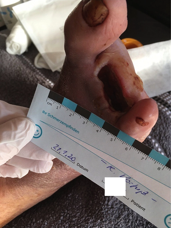
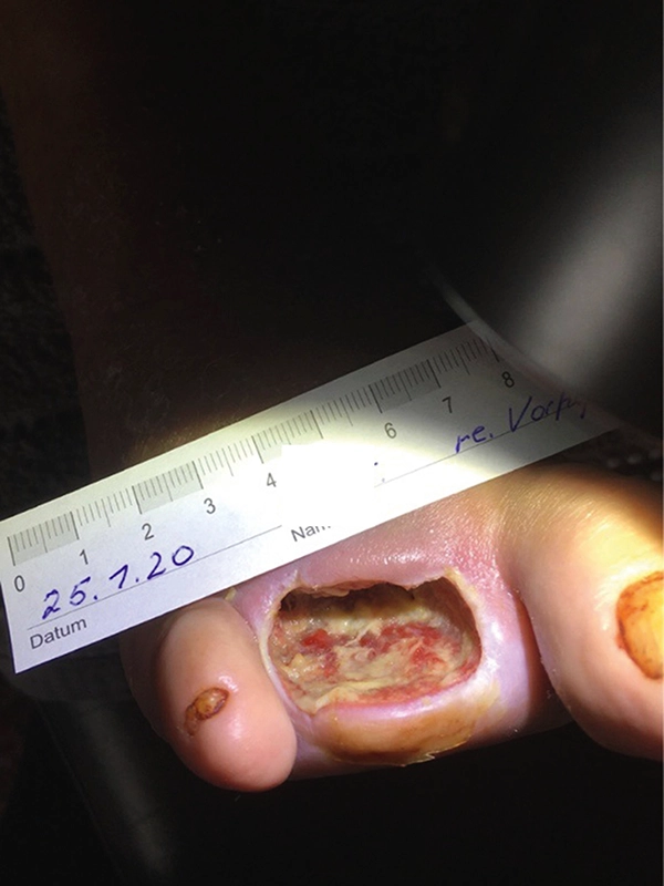
Condition after toe amputation of the second to fourth toes on the right foot shows that 80% of the wound surface was covered with fibrin, with a wound size of 0.6 x 3.5 x 1.6 in (1.5 x 9.0 x 4.0 cm) on January 21, 2020. Wound cleansing and treatment with LIGASANO® began on January 25, 2020. The patient was initially very unhappy with the prognosis for wound healing, which was projected for summer 2021.
-
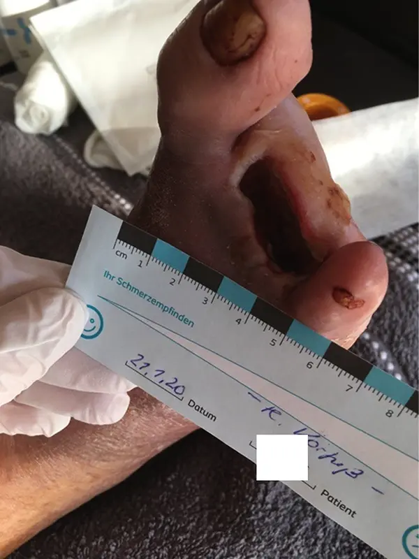
Fig. 1: on January 21, 2020, the condition before starting therapy with LIGASANO® showed a wound size of 0.6 x 1.2 x 1.6 in (1.5 x 3.0 x 4.0 cm), with 80% of the wound covered with fibrin. -
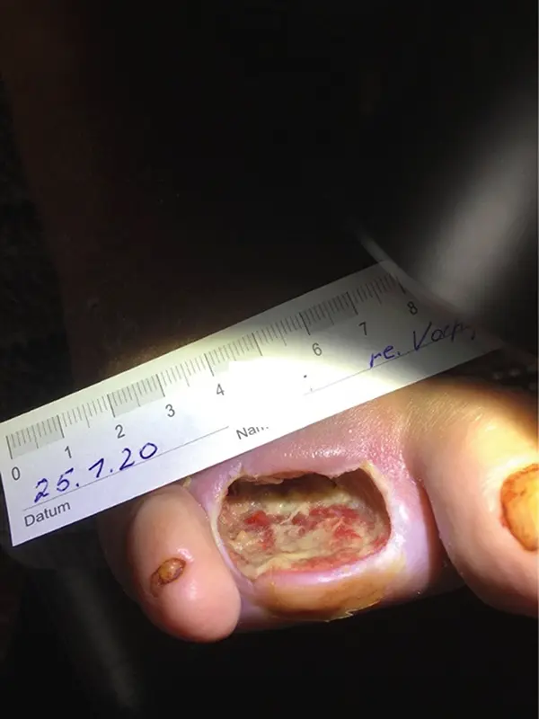
Fig. 2: on January 25, 2020, the wound size was 0.8 x 1.2 x 1.6 in (2.0 x 3.0 x 4.0 cm), with 60% of the wound covered with fibrin. -
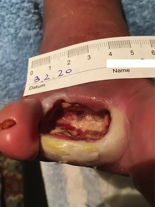
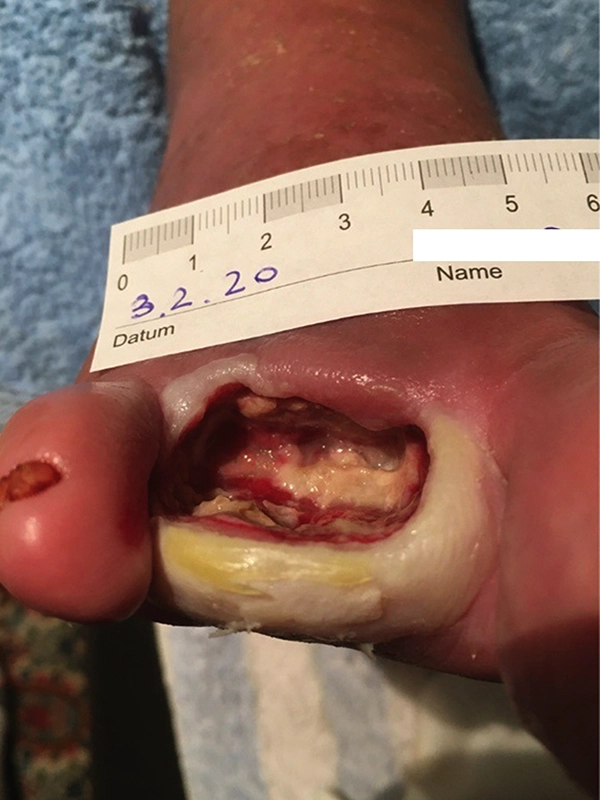
Fig. 3: on February 3, 2020, the wound size was 1.0 x 1.2 x 1.6 in (2.5 x 3.0 x 4.0 cm), with 40–50% of the wound covered with fibrin and moist necrosis at the wound edge. -
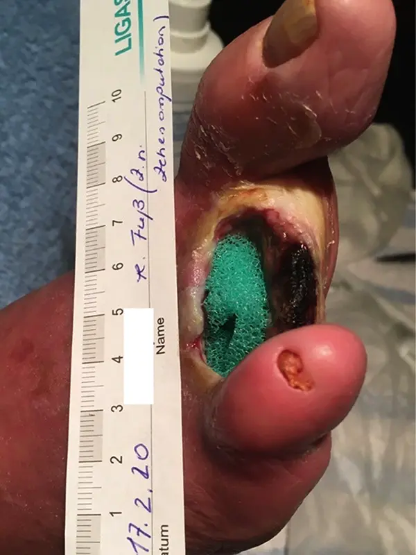
Fig. 5: on February 24, 2020, the wound size was 0.9 x 1.2 x 1.2 in (2.3 x 3.0 x 3.0 cm), with 60% of the wound covered with fibrin and moist necrosis at the wound edge. -
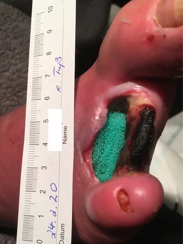
Fig. 4: on February 17, 2020, the wound size was 0.9 x 1.2 x 1.4 in (2.3 x 3.0 x 3.5 cm), with 60–70% of the wound covered with fibrin and moist necrosis at the wound edge. -
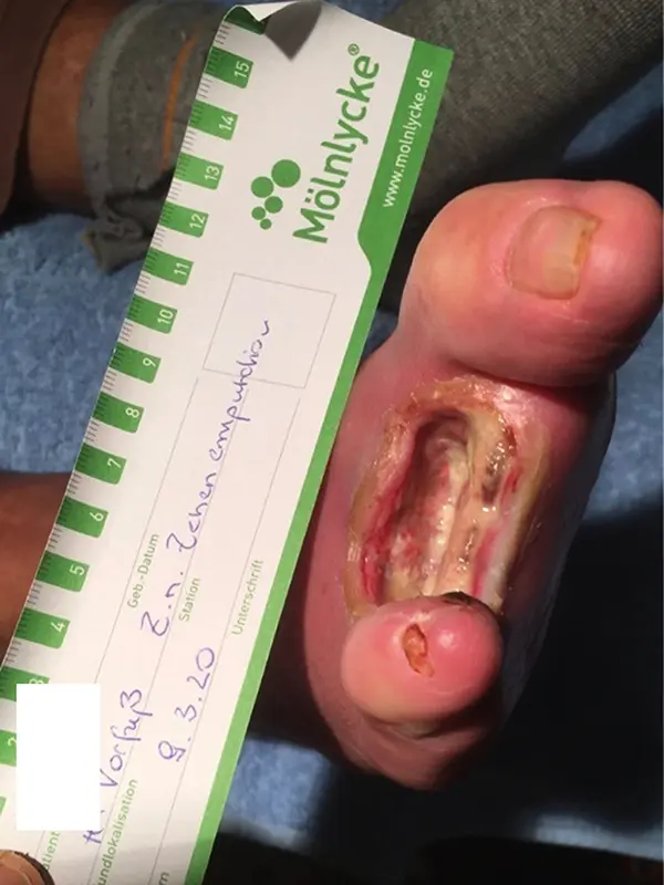
Fig. 6: on March 9, 2020, the wound size was 0.8 x 1.2 x 1.1 in (2.0 x 3.0 x 2.8 cm), with 50% of the wound covered with fibrin and moist necrosis at the wound edge. -
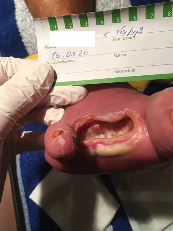
Fig. 7: on March 16, 2020, the wound size was 0.8 x 1.2 x 1.0 in (2.0 x 3.0 x 2.5 cm), with 30–40% of the wound covered with fibrin and moist necrosis at the wound edge. -
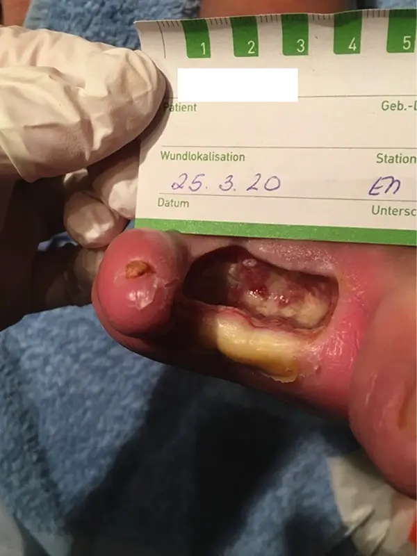
Fig. 8: March 25, 2020: decrease in exudation; the wound is now covered with approximately 30% fibrin.
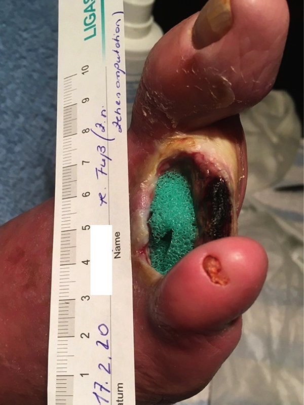
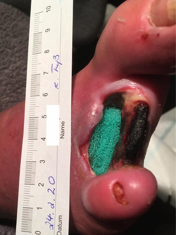
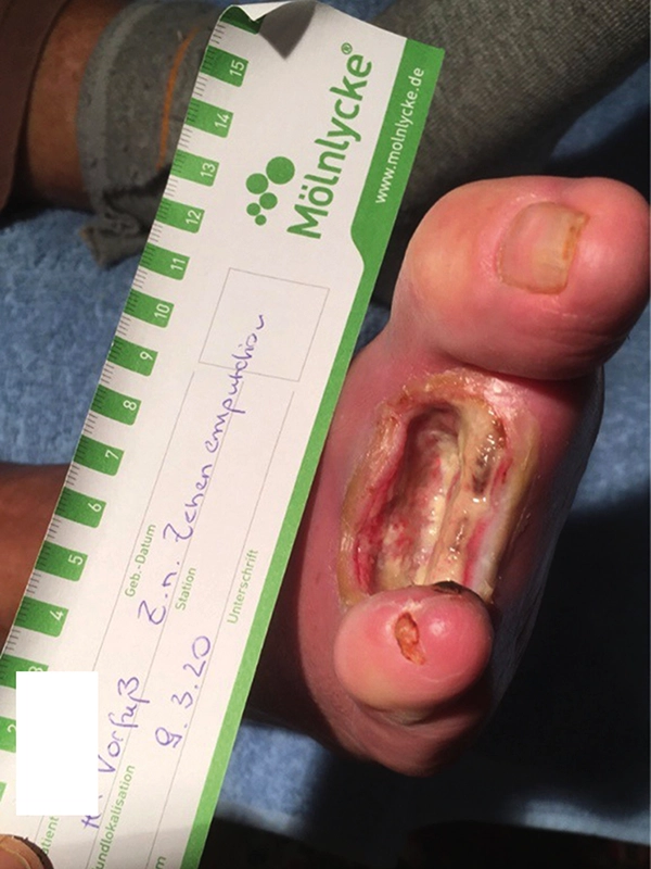
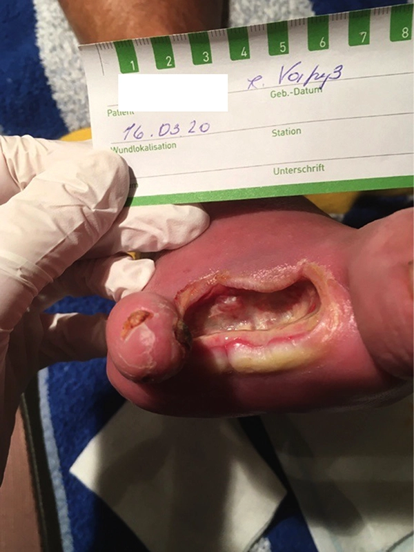
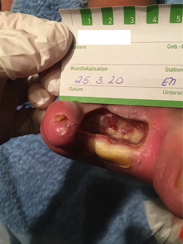
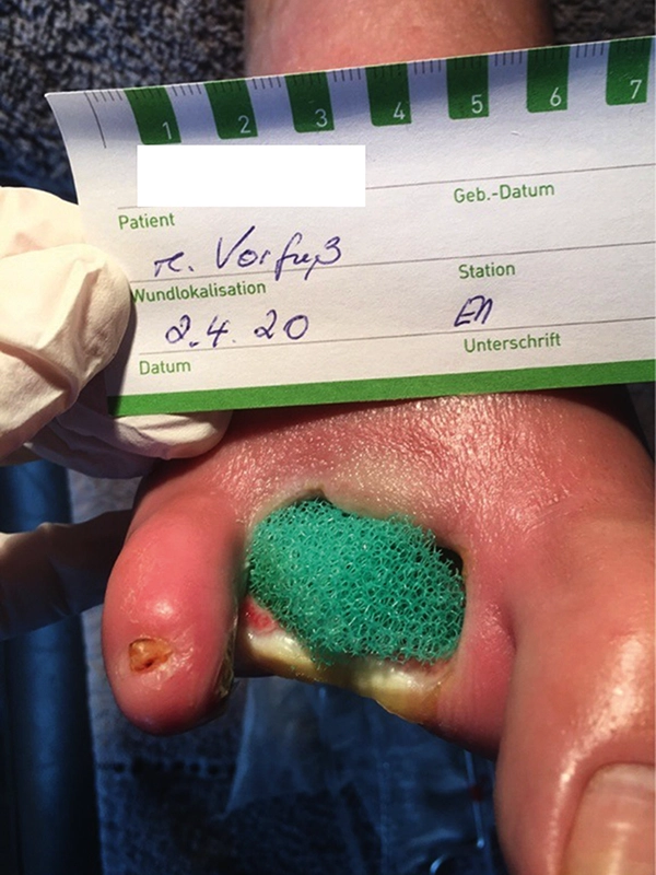
LIGASANO® Green Scrubber was used for mechanical debridement. The wound cavity was then packed with LIGASANO® Dressing Foam.
-
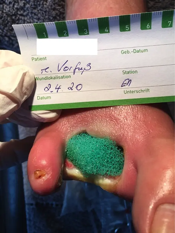
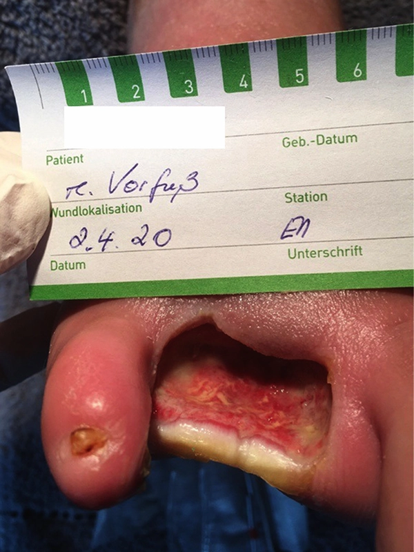
Fig. 9: April 2, 2020: Wound cleansing with LIGASANO® Green Scrubber to remove the moist necrosis at the wound edge. -
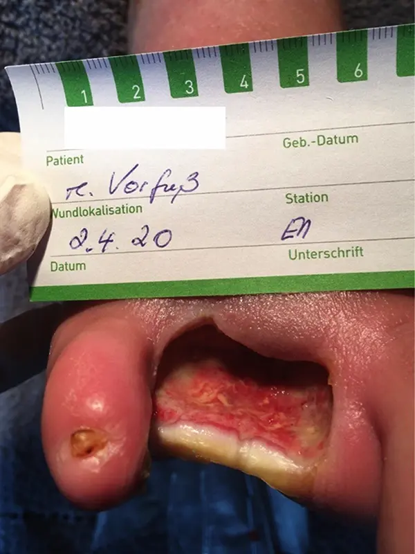
Fig. 10: April 2, 2020: Wound size 0.8 x 1.2 x 0.8 in (2.0 x 3.0 x 2.0 cm), with 10–20% of the wound covered with fibrin; wound care with LIGASANO® White Dressing. -
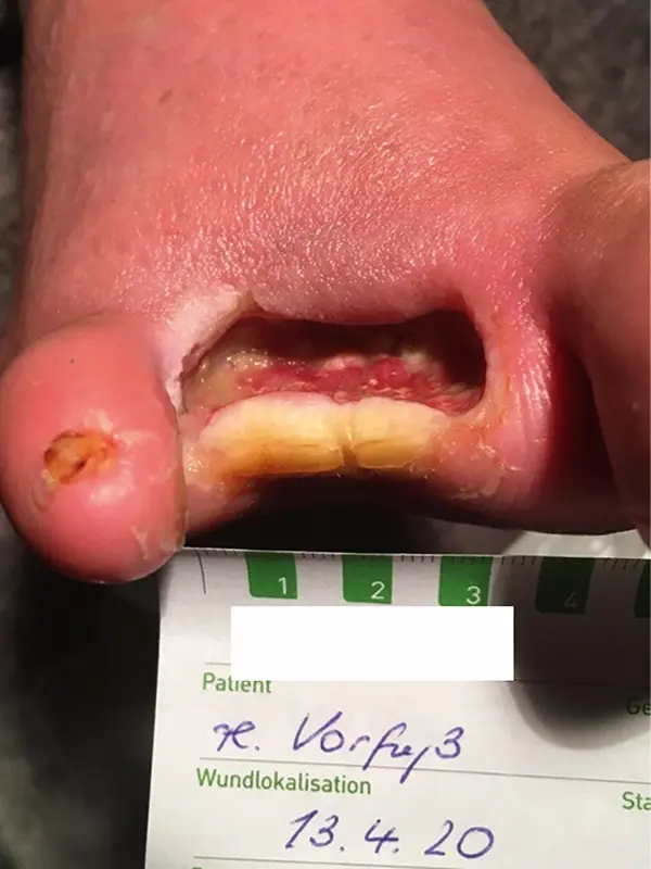
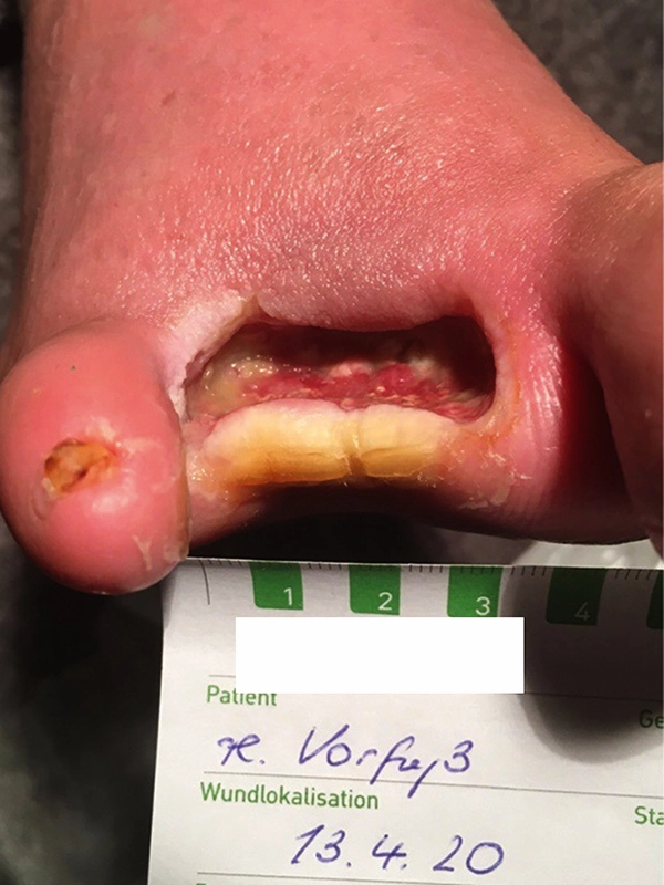
Fig. 11: April 13, 2020 wound size 0.7 x 1.2 x 0.8 in (1.8 x 2.8 x 2.0 cm). -
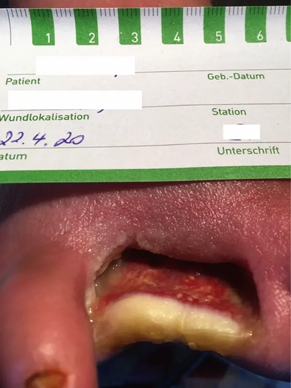
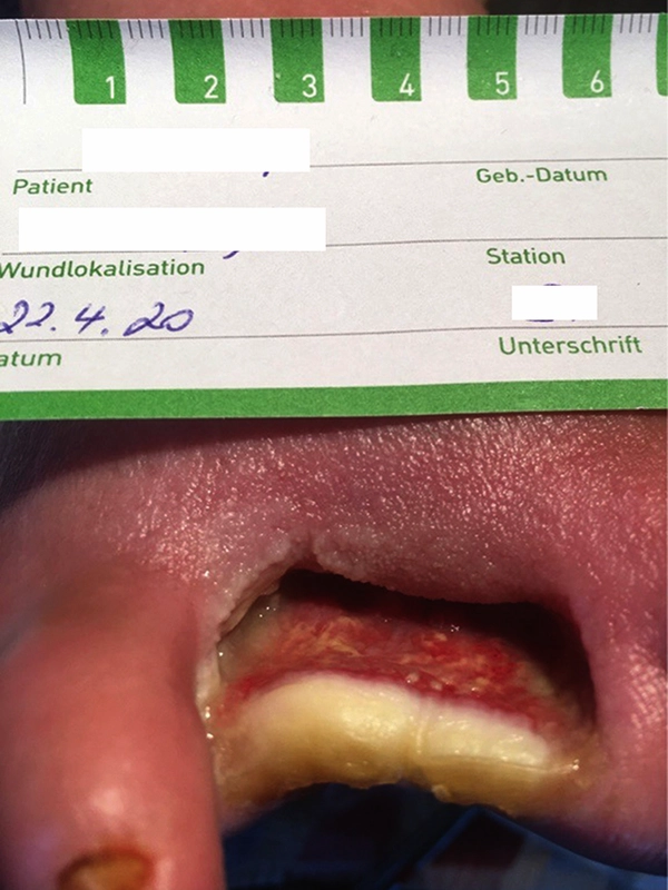
Fig. 12: April 22, 2020: wound still approximately 10% covered with fibrin; yellow necrosis present at the upper wound edge. -
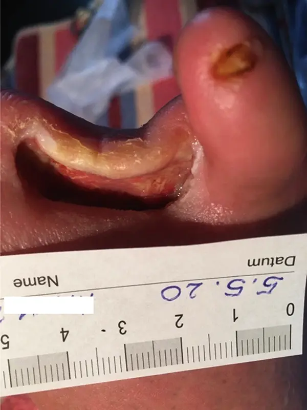
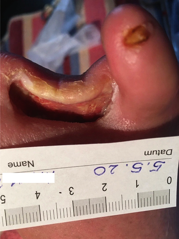
Fig. 13: May 5, 2020: wound barely exuding; LIGASANO® White Dressing moistened with hydrogel. -
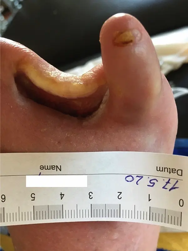
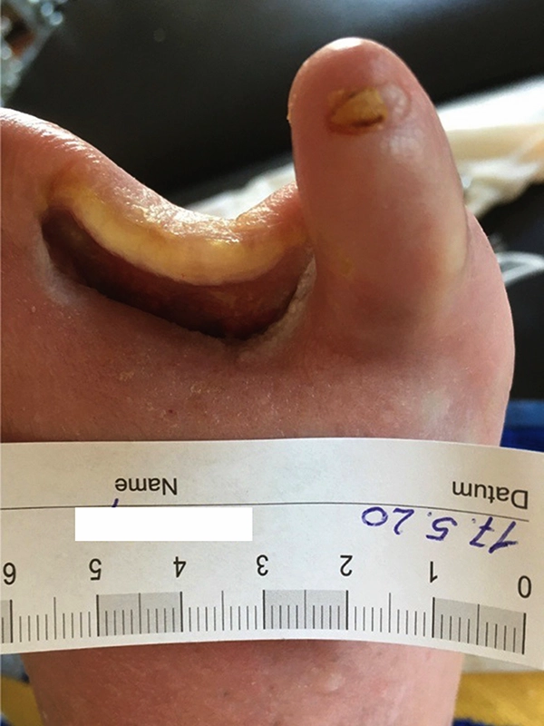
Fig. 14: May 17, 2020: wound size 0.6 x 0.8 x 0.6 in (1.5 x 2.0 x 1.5 cm), no more fibrin deposits present. -
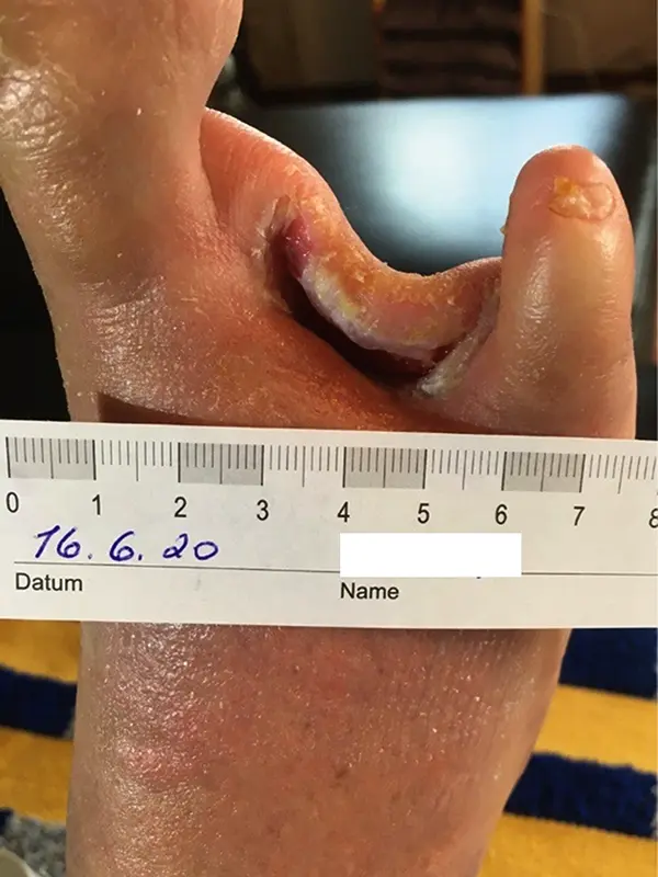
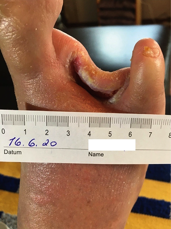
Fig. 16: June 16, 2020: wound size 0.4 x 0.7 x 0.4 in (1.0 x 1.8 x 1.0 cm). Wound-edge coating had decreased. -
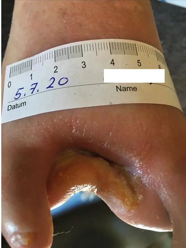
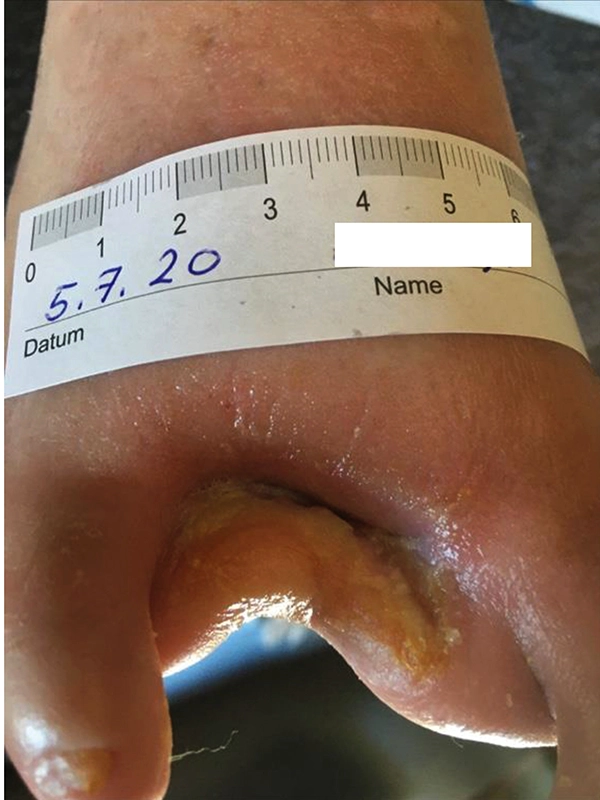
Fig. 17: July 5, 2020: wound size 0.3 x 0.4 x 0.2 in (0.8 x 1.0 x 0.5 cm). There is a relatively rapid increase in epithelialization. -
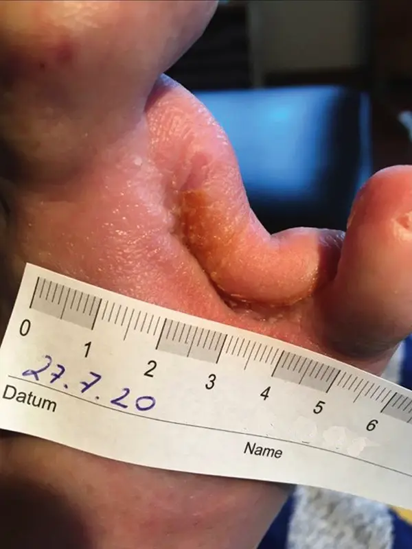
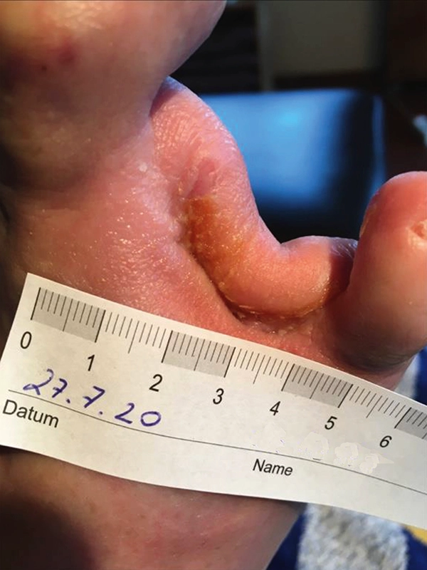
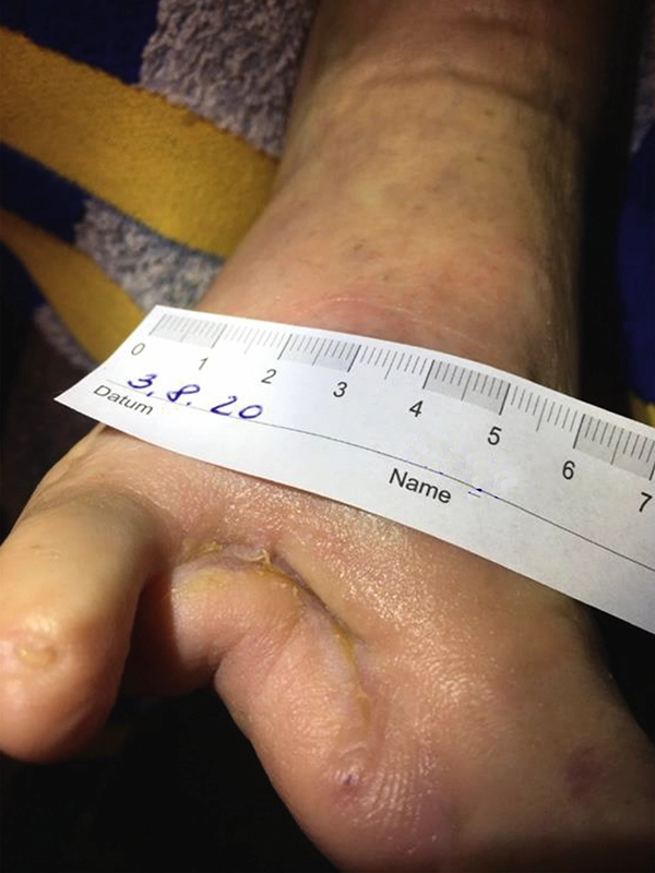
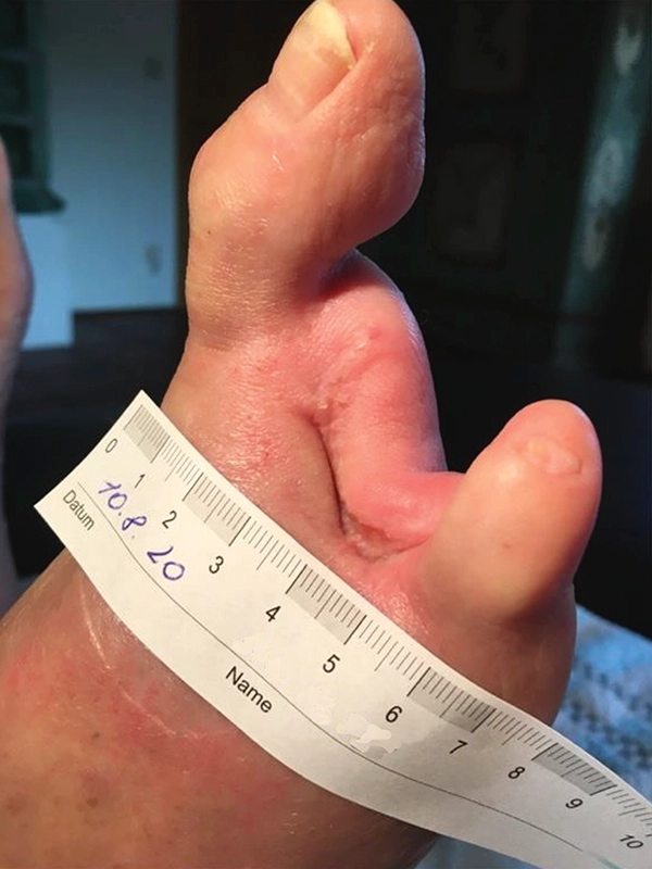
Fig. 18: July 27, 2020: the wound healing is complete. -
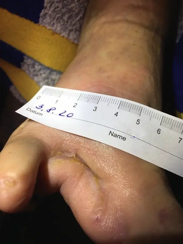
Fig. 19: August 3, 2020 -
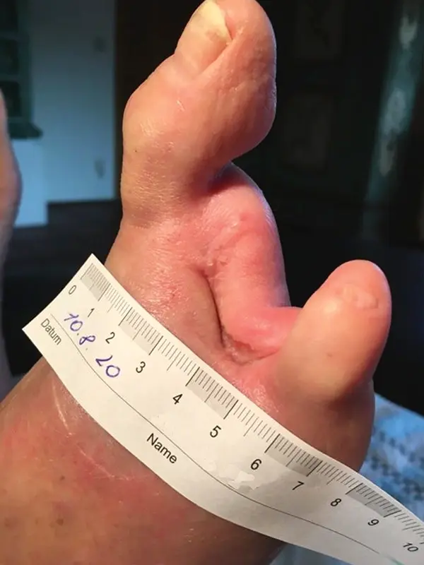
Fig. 20: August 10, 2020
Conclusion:
Complete epithelialization was documented on July 27, 2020. The patient is greatly relieved that his wound has now healed, marking a return to normalcy and an improvement in his quality of life. The scar will be treated with calcium fluoratum ointment until further notice. Additionally, the patient now has orthopedic shoes.