
Oktober 2019 | Practical Experience Report by Cristina Krause-Mohr, Wound Specialist (ICW), Owner of the Nursing Service Krause in Nickenich, Germany
Patient Data and Medical History:
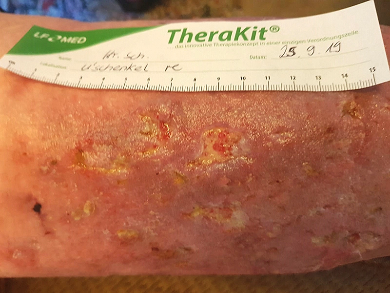
A 65-year-old male patient with type 2 diabetes, obesity, and chronic venous insufficiency (CVI) on both sides had developed multiple wounds on his legs over several years. His mobility was limited. Due to personal reasons, he initially refused inpatient treatment and relied on outpatient vascular consultations and local nursing services. Despite a year of treatment with zinc paste dressings, there was no significant improvement. Eventually, the patient agreed to inpatient care, which was conducted at Heilig Geist Hospital in Boppard under the care of PD Dr. Gunnar Riepe. The inpatient stay lasted 14 days and included lymphatic drainage, physiotherapy, and the initiation of treatment with LIGASANO® White Dressing. The first visit was on September 3, 2019.
Anwenderbericht
-
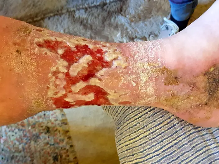
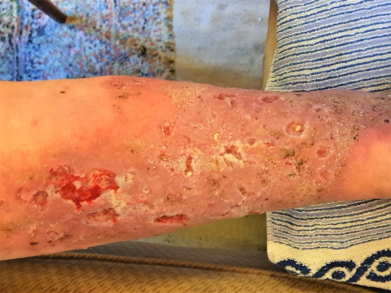
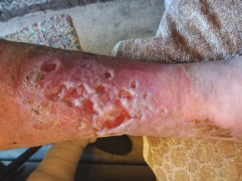
Fig. 1: on September 3, 2019, the condition of the left lower leg showed a large wound with several areas of macerated tissue and a macerated wound edge. The wound exuded moderately, with the wound bed showing good blood supply and some fibrin presence. The surrounding skin was dry and scaly. -
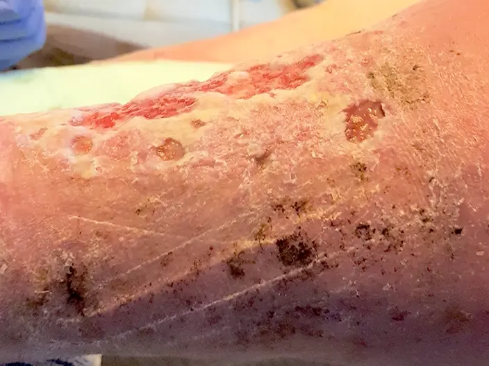
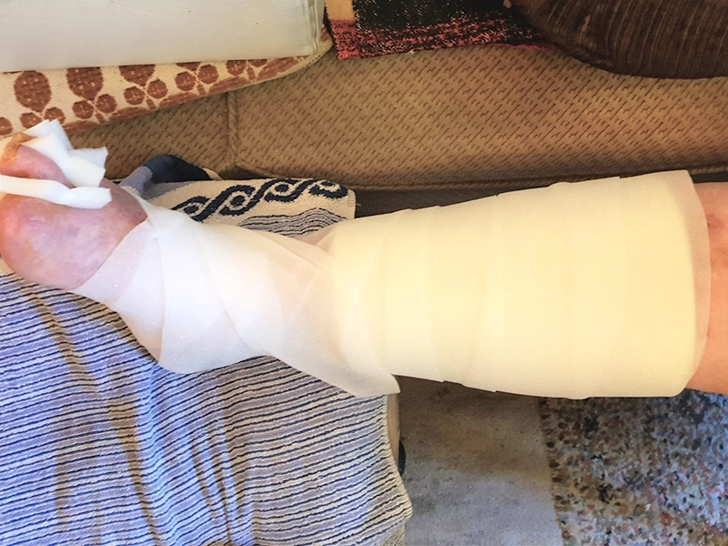
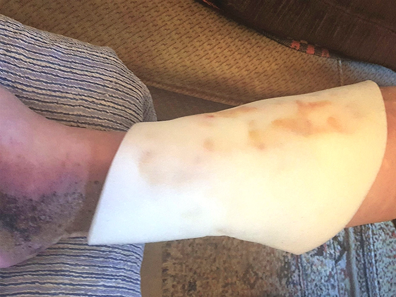
Fig. 2: on September 3, 2019, for the outer side of the left lower leg, the wound was cleansed with sodium chloride 0.9%. A LIGASANO® White Dressing, 9.5 x 6.3 x 0.4 in (24 x 16 x 1 cm) in size, was applied to the wound and surrounding area. For fixation, a bandage of LIGASANO® Pressure Relief Foam, measuring 118 x 4 x 0.12 in (300 x 10 x 0.3 cm), was used. The lower leg was then wrapped with short-stretch bandages. -
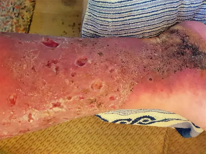
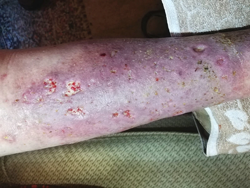
Fig. 3: on September 3, 2019, the right lower leg showed several wounds of varying depths with furrowed and partially macerated edges. The surrounding skin was very dry and scaly. -
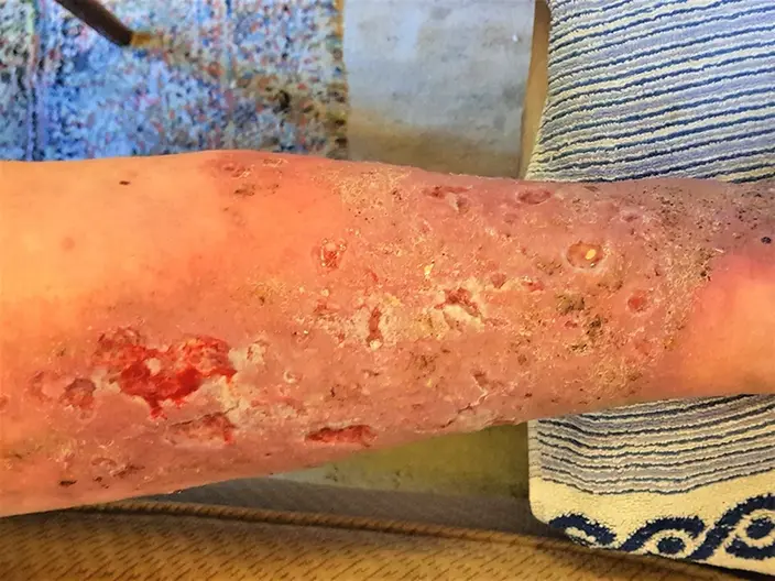
Fig. 4: on September 3, 2019, after cleaning the wound with sodium chloride 0.9%, LIGASANO® White Dressing, measuring 11.6 x 9.6 x 0.4 in (29.5 x 24.5 x 1 cm), was applied. This dressing covered almost the entire lower leg, including the wounds and surrounding skin. It was secured with a bandage of LIGASANO® Pressure Relief Foam, 10 cm wide. -
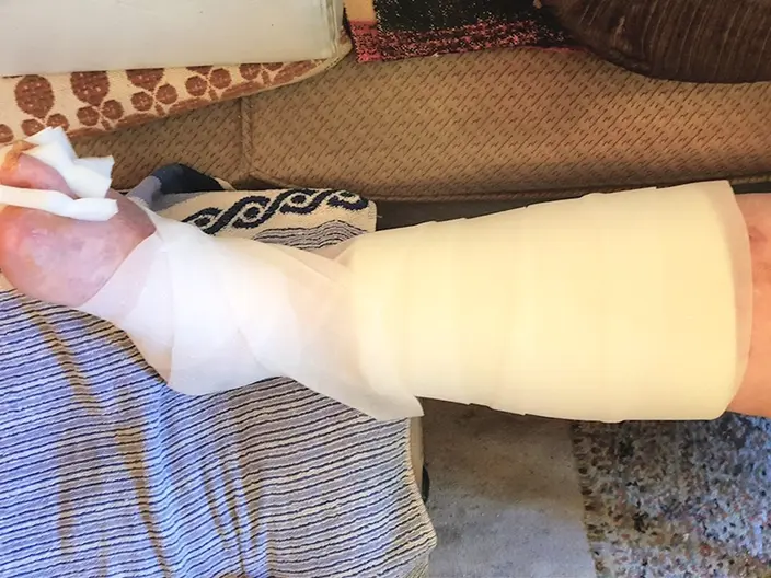
Fig. 5: on September 3, 2019, a compression bandage was applied to the right lower leg using short-stretch bandages. The Krause nursing service from Nickenich changed the dressing daily. -
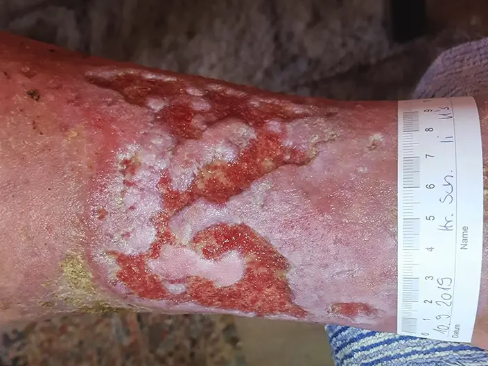
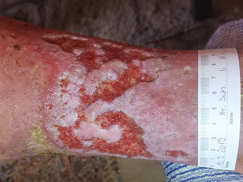
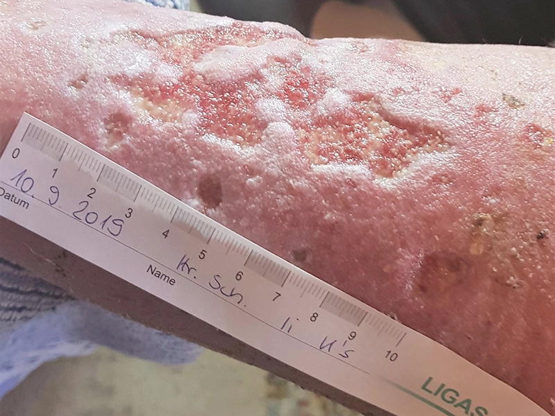
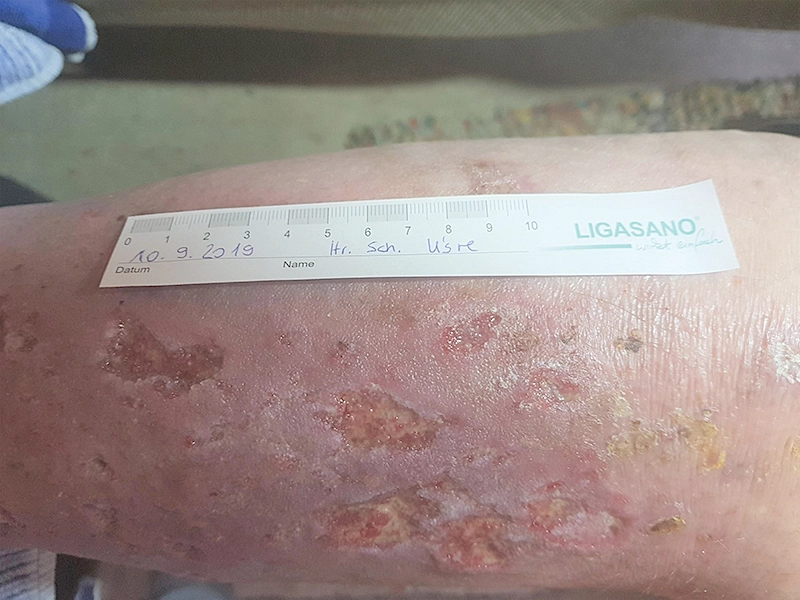
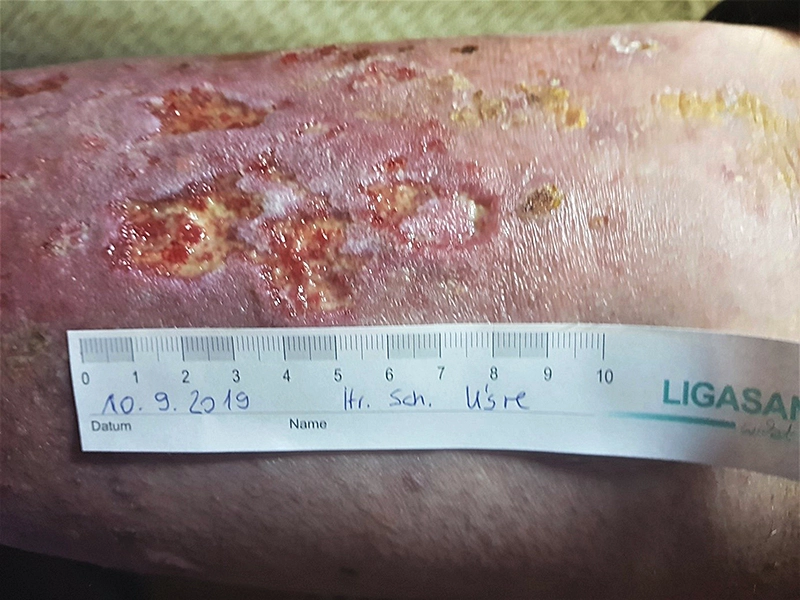
Fig. 6: on September 10, 2019, the left lower leg showed significant improvement. Tissue islands and wound edges were no longer macerated, and the wound bed was well perfused. The condition of the wound environment had improved, with significantly fewer dry skin flakes present. -
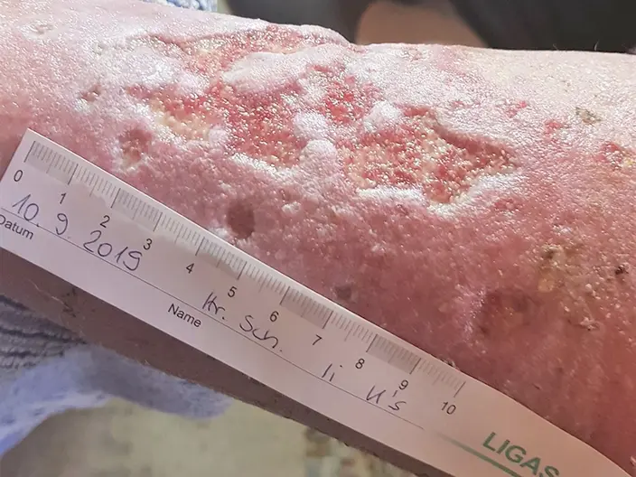
Fig. 7: the lower left leg on September 10, 2019. -
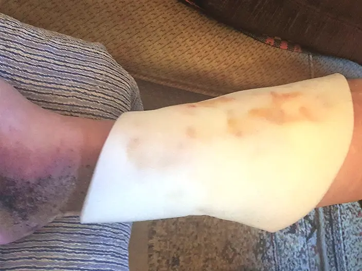
Fig. 8: September 10, 2019: LIGASANO® White Dressing covering the lower right leg before the dressing change. -
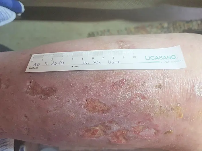
Fig. 9: on September 10, 2019, significant improvement was also visible on the right lower leg. The wound edges were no longer macerated, though the wound bed was still partially covered with fibrin. The surrounding skin was less dry, with hardly any skin flakes present. -
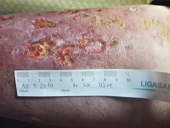
Fig. 10: on September 10, 2019, the treatment for the right lower leg wound continued without changes, except that wound cleansing was switched from sodium chloride 0.9% to Prontosan wound irrigation solution. -
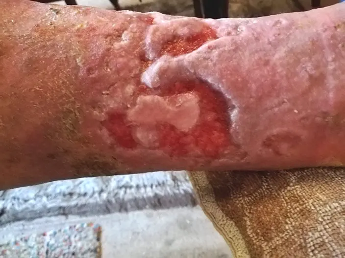
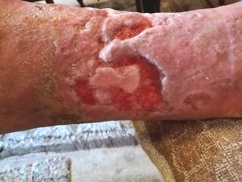
Fig. 11: on September 18, 2019, the left lower leg showed a significant reduction in wound size. Granulation was progressing well, the wound bed was well supplied with blood, and the surrounding skin had become partially drier again. -
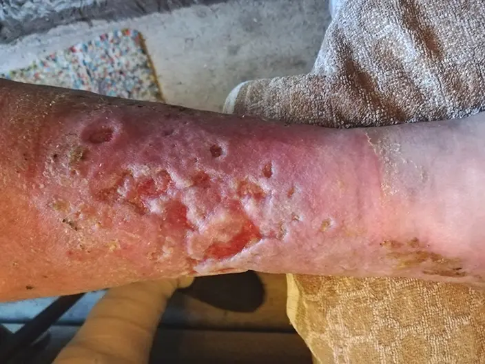
Fig. 12: the lower left leg on September 18, 2019. -
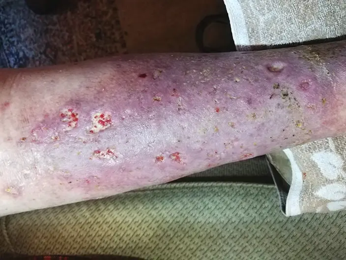
Fig. 13: on September 18, 2019, the right lower leg still had some partial fibrin deposits. The treatment continued as previously described. -
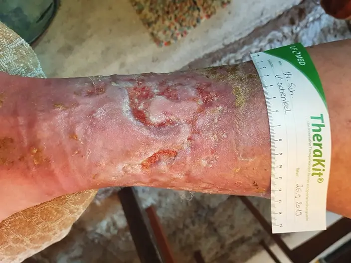
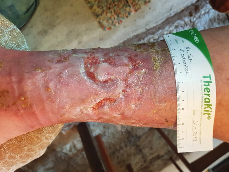
Fig. 14: on September 26, 2019, the left lower leg was treated with LIGASANO® White Dressing, size 5.9 x 4.0 x 0.4 in (15 x 10 x 1 cm), and a bandage of LIGASANO® Pressure Relief Foam. -
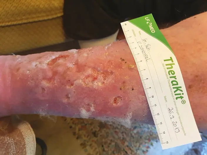
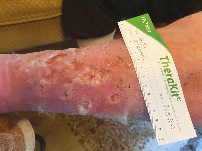
Fig. 15: September 26, 2019 -
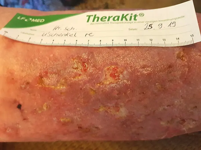
Fig. 16: on September 26, 2019, the right lower leg showed partial fibrin deposits. A hydrogel was used, with a thin layer of Prontosan wound gel applied to the fibrin-covered areas. The LIGASANO® White Dressing was then applied and secured with a bandage of LIGASANO® Pressure Relief Foam. Compression bandages were also applied using a short-stretch bandage. -
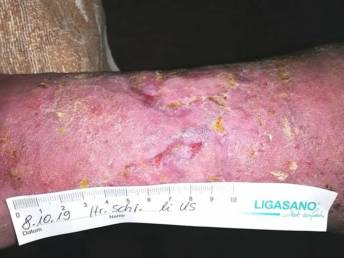
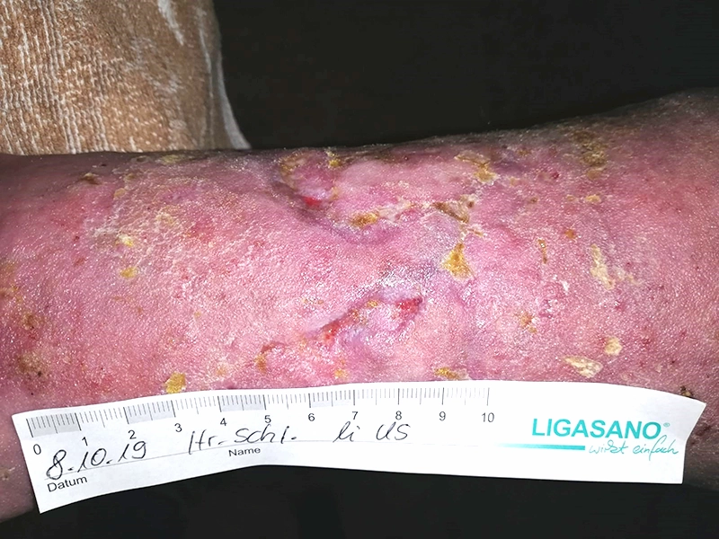
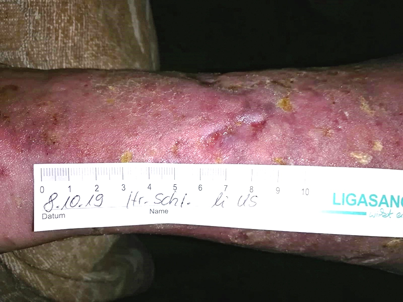
Fig. 17: on October 8, 2019, the wound on the left lower leg was almost closed. To protect the newly formed tissue, a LIGASANO® White Dressing was applied. Skin care was performed using the patient‘s own care lotion. -
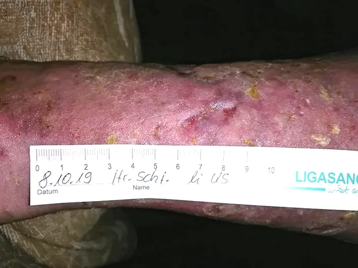
Fig. 18: on October 8, 2019, for the left lower leg, compression therapy was continued using a short-stretch bandage, with padding provided by a bandage of LIGASANO® Pressure Relief Foam. -
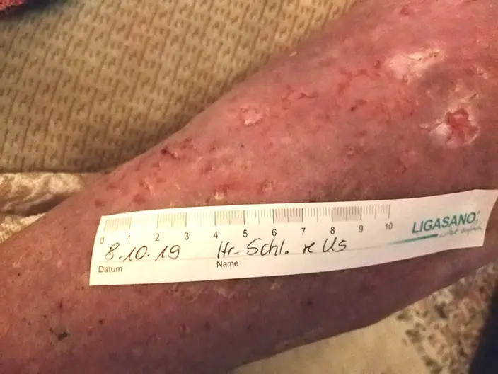
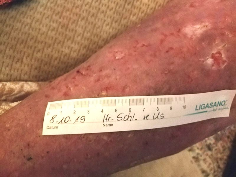
Fig. 19: on October 8, 2019, for the right lower leg, fibrin deposits had detached and granulation was progressing. After cleansing the wound with Prontosan wound irrigation solution, treatment included a LIGASANO® White Dressing and a bandage of LIGASANO® Pressure Relief Foam. Compression was applied using a medium short-stretch bandage. -
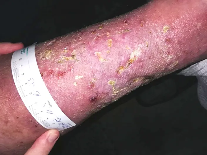
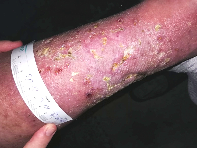
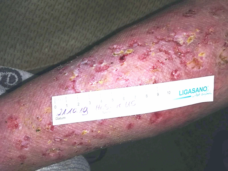
Fig. 21: the lower right leg on October 21, 2019. -
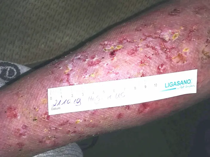
Fig. 20: the lower left leg on October 21, 2019.
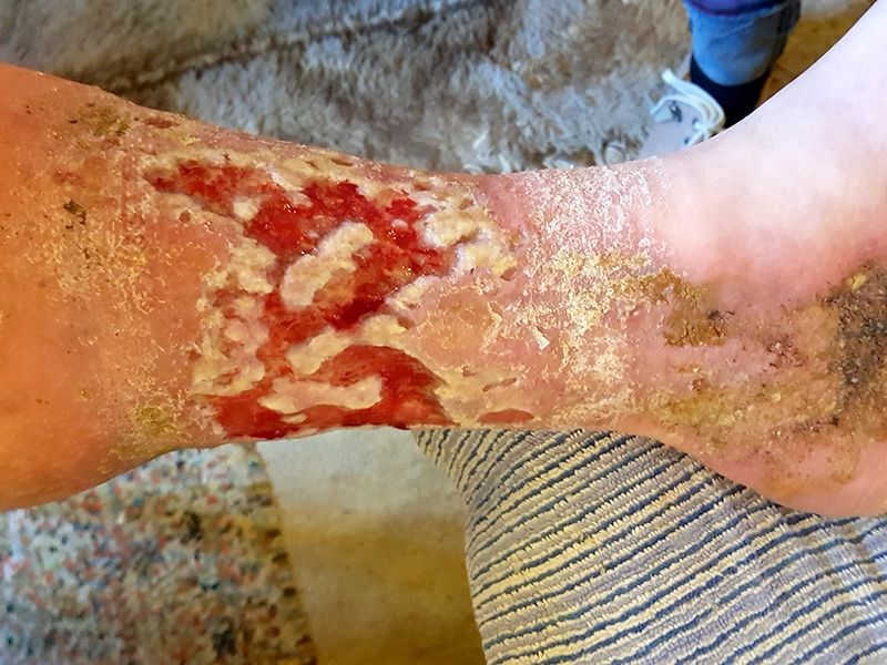
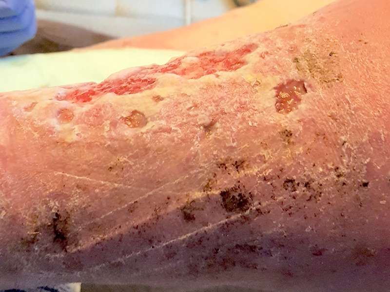
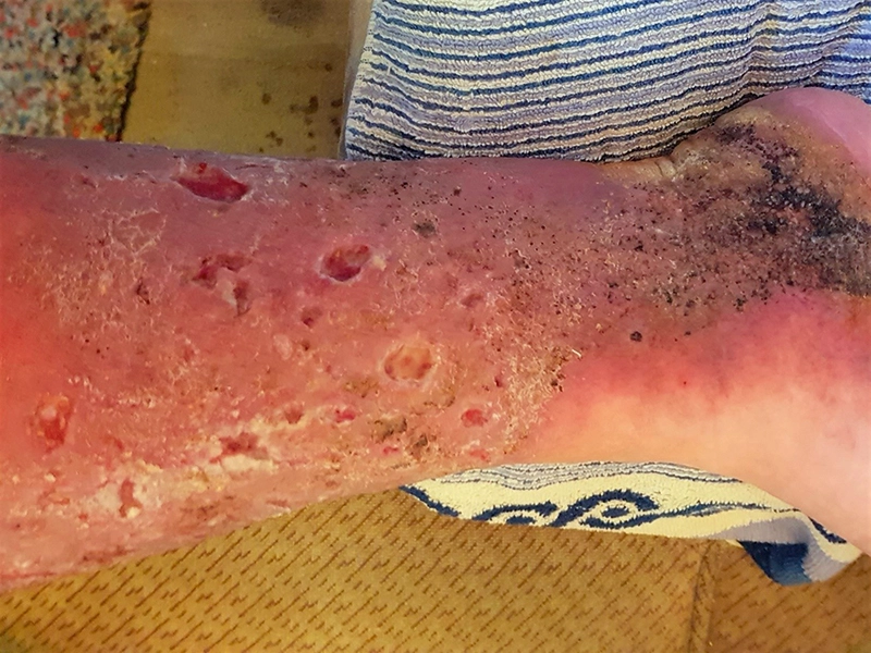
On October 16, 2019, the patient returned to Heilig Geist Hospital in Boppard for Dr. Riepe’s wound consultation due to plantar wounds. Treatment included a TCC (Total Contact Cast).