
February 2016 | Practical Experience Report by Heidi Jodl, Nurse, Wound Manager AZWM and Head of the WZ® Wound Center Augsburg, Germany
Patient Data and Medical History:
A 52-year-old male patient with diabetic foot syndrome had a forefoot amputation on the left side in February 2016. His medical history includes PAD grade 2 on the left, insulin-dependent type 2 diabetes mellitus, polyneuropathy, chronic renal insufficiency following acute renal failure, and a history of alcohol abuse. Additionally, he suffers from hypokalemia, hypocalcemia, hypertensive diabetic nephropathy, and left MCA stroke. Other conditions include arterial hypertension, hyperlipidemia, and nicotine abuse.
Despite hemiparesis on the right side, the patient is mobile with a forefoot offloading shoe with a continuous sole and a compensating sole on the right side. He spent 60 days in inpatient care. Due to the formation of a 1.6 in (4 cm) deep fistula in the medial forefoot area, another amputation or a lower leg amputation was suggested. Concerned about the prospect of extended hospitalization, the patient was transferred to the WZ® Wound Center Augsburg in April. During his hospital stay, the critical colonization was managed with an antiseptic containing octenidine dihydrochloride and 2.0 g phenoxyethanol (Octenisept®) for wound cleaning. The wound was treated with a silver-containing hydrofiber as the filler and an absorbent dressing for coverage, with daily dressing changes due to strong exudation, wound odor, and critical colonization.
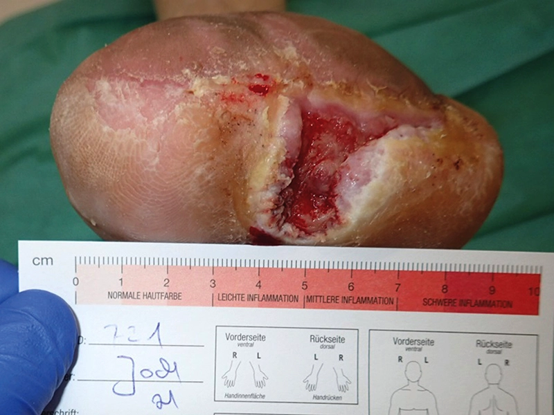
Upon admission to the WZ® Wound Center, the wound environment was edematous, reddened, and slightly overheated. Hyperkeratosis was strongly present at the wound margins, particularly spongy in the plantar area. Removal of the spongy hyperkeratosis revealed a fetid odor. The wound bed showed granulating tissue covered with fibrin. The granulation tissue appeared granular but light pink after the fibrin coating was removed. There was a strong, viscous, cloudy exudation. The local therapeutic wound treatment was initially implemented as follows:
Wound cleaning: The wet-dry phase was performed using a wound cleaning solution based on singlet oxygen (NaOCl) and seawater (ActiMaris®). For skin care and to treat the wound environment, a lotion containing water/oil (w/o) with a lipid content of 27% (BL® - BasicLotion) was applied.
Wound filler: a silver-containing hydrofiber with ethylenediaminetetraacetate (Aquacel® Ag + ExtraTM) was used.
Wound coverage: the wound was covered with Zetuvit® plus 4 x 8 in (10 x 20 cm), and fixation was achieved with an elastic bandage with cotton content, combined with a longitudinal and transverse elastic bandage (Tubifast™ blue 2-Way Stretch Technology®).
Dressing changes were performed daily by the nursing service, and a weekly check-up was conducted at the WZ® Wound Center.
-
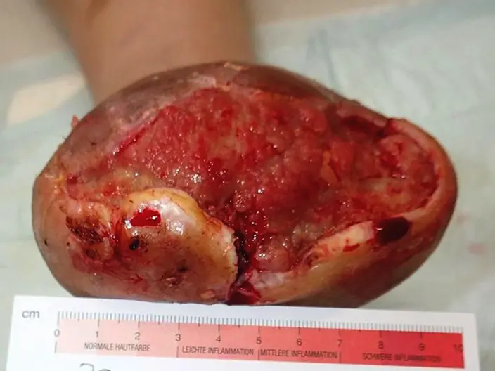
Fig. 1: on April 18, 2016, the forefoot amputation wound on the left side was cleaned and underwent surgical debridement using a curette. -
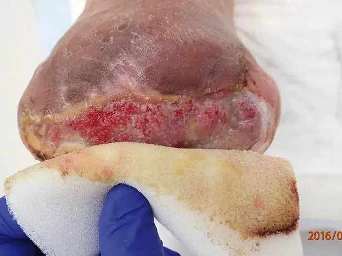
Fig. 2: -
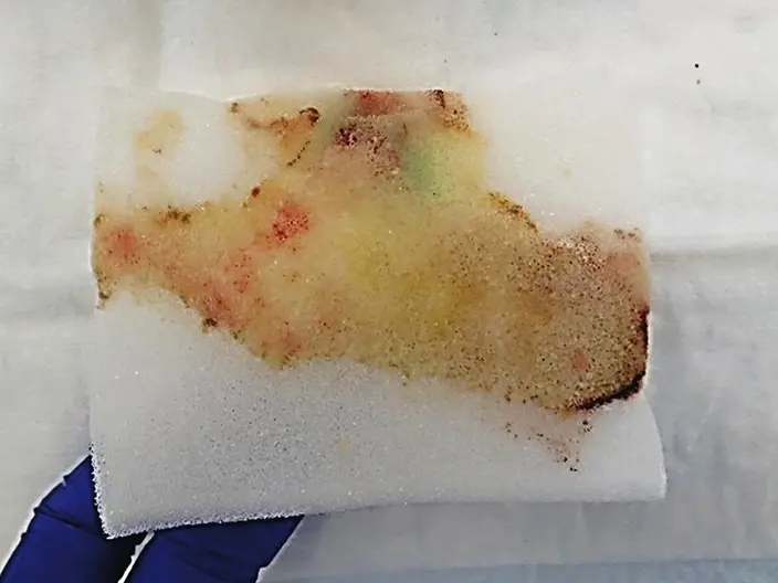
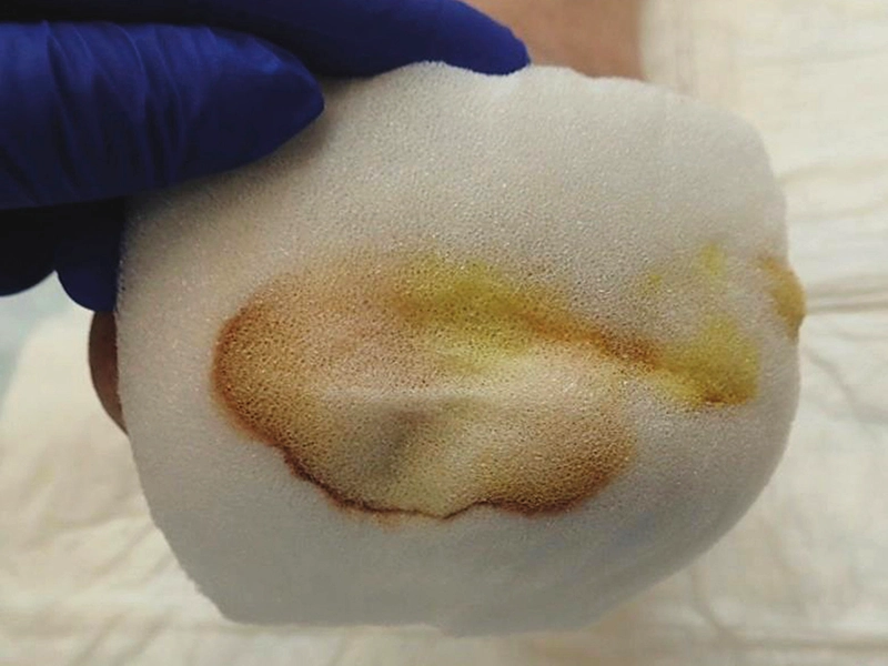
Fig. 2 + 3: on April 25, 2016, during the dressing removal, it was noted that the viscous exudate had been effectively absorbed by LIGASANO® White Dressing without drying out the wound.
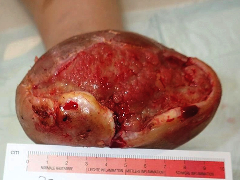
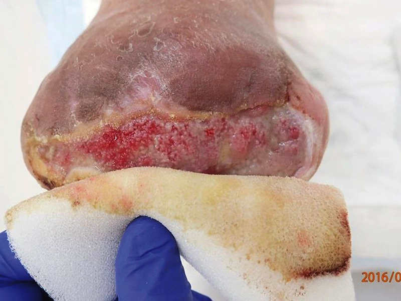
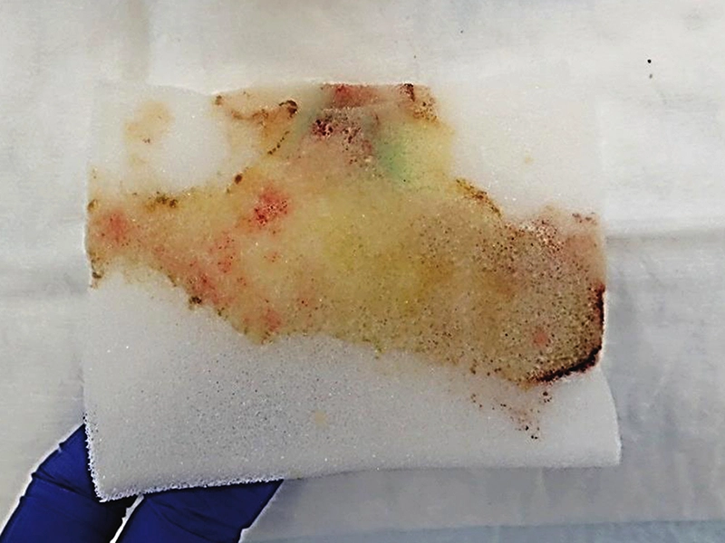
After one week, a viscous exudate appeared beneath the Aquacel® Ag + extra™ when the dressing was removed. Due to its thick consistency, it remained between the wound base and the hydrofiber, which could only absorb it to a limited extent. This exudate congestion inhibited granulation tissue growth. Additionally, the discharge of the exudate from the fistula was limited, showing signs of retention. Therefore, the treatment was switched to LIGASANO® White Dressing, sterile (6 x 4 x 0.4 in / 15 x 10 x 1 cm). The fistula was loosely padded dry with a LIGASANO® White Dressing for cavities (39.4 x 0.6 x 0.16 in / 100 x 1.5 x 0.4 cm).
Under continuous local therapeutic measures using the LIGASANO® White Dressing (size 39.4 x 0.6 x 0.16 in resp. 100 x 1.5 x 0.4 cm), the fistula closed, and the amount of exudation was significantly reduced. The wound bed displayed well-perfused, granular granulation tissue with no detectable odor. Additionally, there was a noticeable reduction in hyperkeratosis and maceration.
-
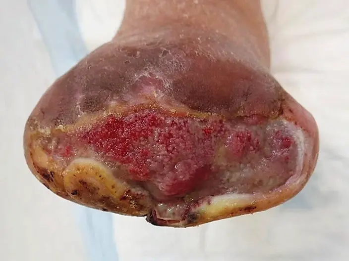
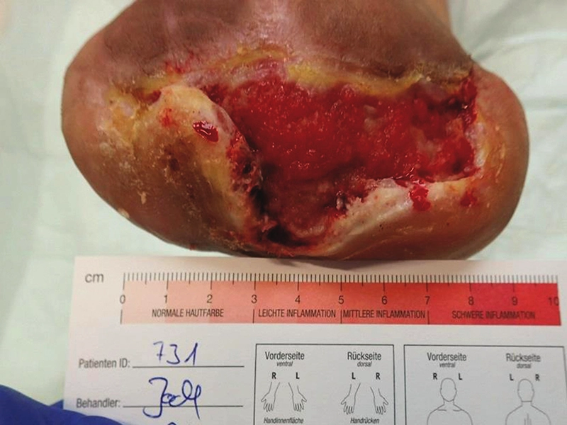
Fig. 4: on April 25, 2016, the wound condition before the wet-dry phase, immediately after the dressing was removed, showed a reduction in hyperkeratosis and fibrin deposition. Granulation tissue had also begun to develop. -
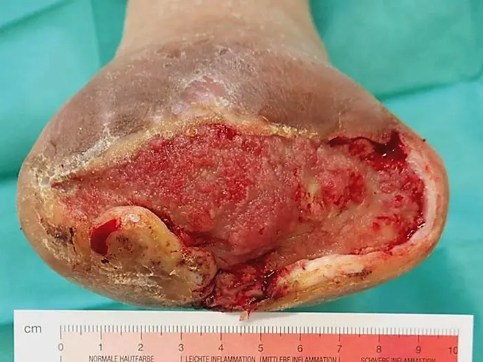
Fig. 5: on April 25, 2016, after the wet-dry phase and surgical debridement, the wound condition showed that the depth of the fistula had been reduced to 0.8 in (2 cm). -
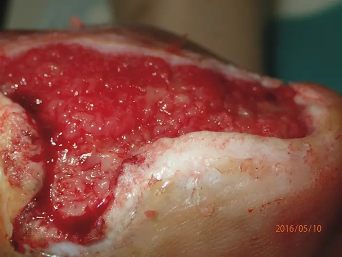
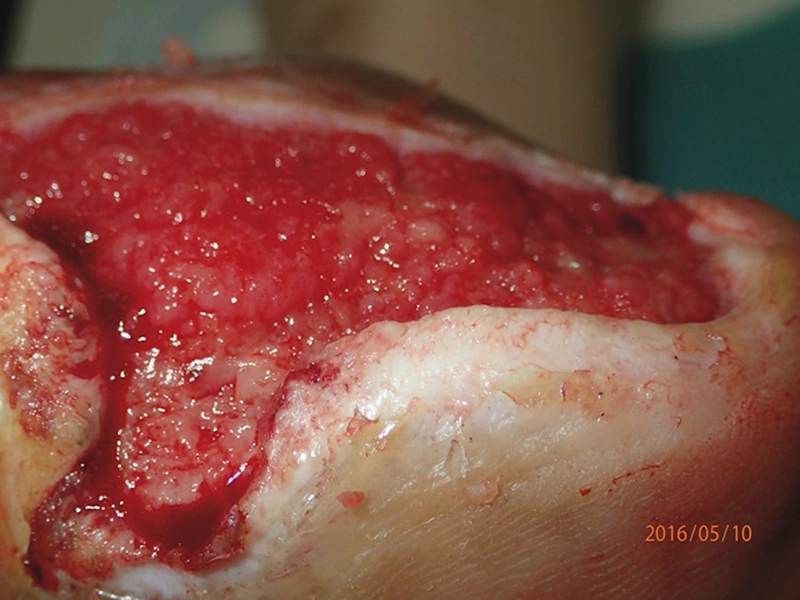
Fig. 6: on May 10, 2016, after the wet-dry phase and cleansing with LIGASANO®, red, granular, and vital granulation tissue was visible. -
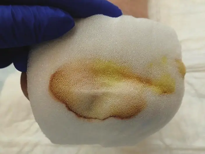
Fig. 7 + 8: wound condition on May 23, 2016 during removal of LIGASANO® White Dressing -
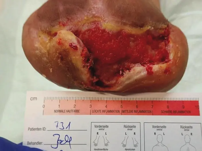
Fig. 8: -
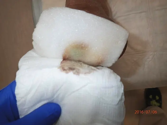
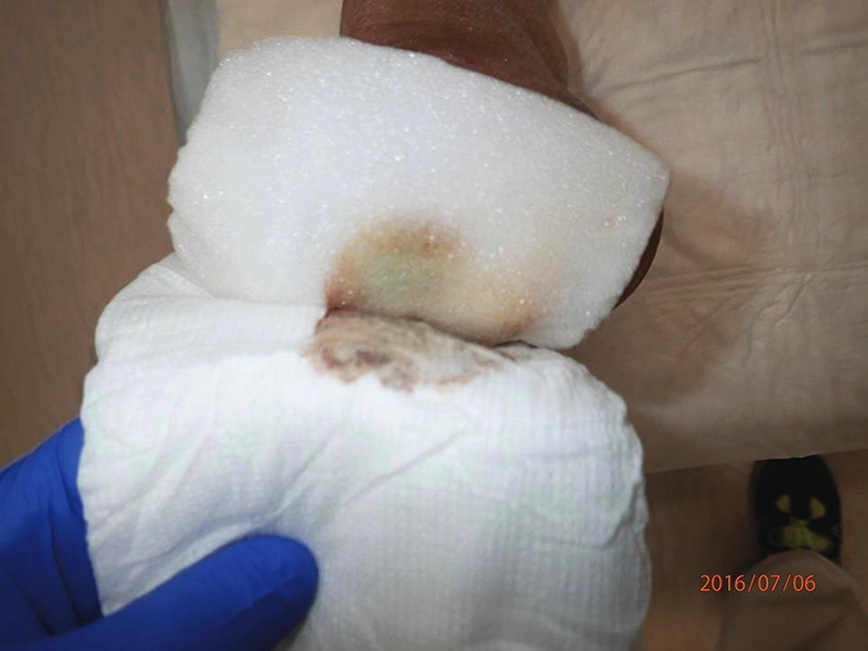
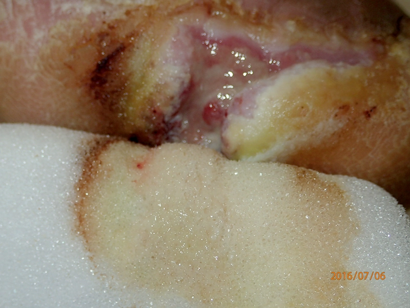
Fig. 9: on July 6, 2016: dressing remov al revealed that the absorbent dressing served as the wound cover, with the vertical diffusion of exudate effectively managed by LIGASANO® Wound Dressing. -
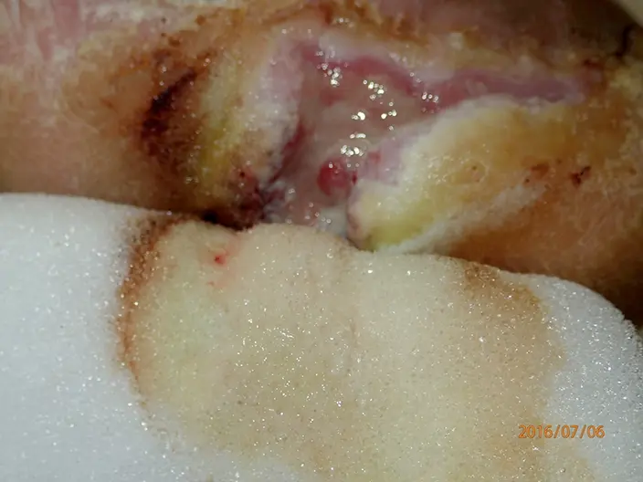
Fig. 10: -
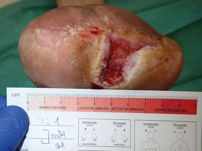
Fig. 10 + 11: on July 6, 2016: the structure and pore openings of LIGASANO® White Dressing were visible, demonstrating its effective absorption of the viscous exudate while simultaneously protecting the wound margins and surrounding area. During the treatment with LIGASANO® White Dressing, the wound environment was stabilized, and the patient‘s well-being was enhanced through targeted exudate management. This included the absorption and vertical diffusion of the viscous exudate, reduction in the amount of exudate, and a decrease in odor.
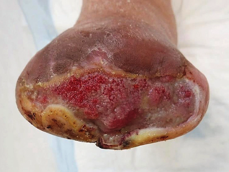
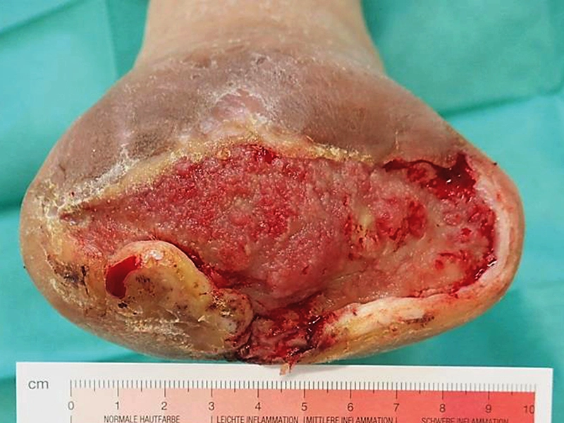
Summary / Conclusion:
The structure of the LIGASANO® White Dressing enabled focused exudate management and controlled absorbency through vertical redirection. Over the course of treatment, fibrin coverage and exudate decreased and granulation tissue was observed.