
May 2018 | Practical Experience Report by Advivo ambulante Beatmung GmbH, Munich, Germany, under the direction of Carola Boser, Nursing Management, and Dr. med. Luis Antonio da Silva Jäger, Munich, Germany
Patient Data and Medical History:
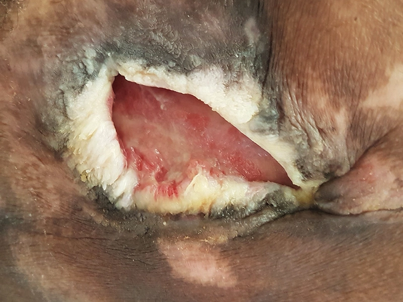
A 53-year-old male patient, dark-skinned, with complete paraplegia below T8 following a work accident in 1996, has experienced recurring pressure ulcer over recent years. He has had a colostomy since December 2015 and underwent plastic surgery in the gluteal region in 2017. His medical history includes anasarca, cardiomyopathy, and type 2 diabetes. The patient presents with an extensive stage 4 pressure injury, that is secondarily infected.
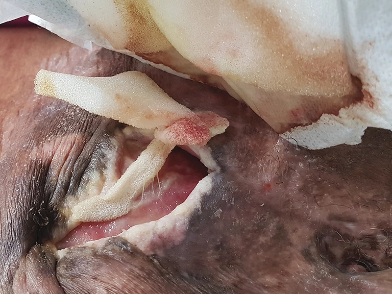
- The wound measures approximately 5.12 in (13 cm) in length, 2.76 in (7 cm) in width, and about 0.4 in (1 cm) in depth, with a 0.08 in (0.2 cm) undermined edge
- The wound edges are heavily macerated
- No signs of inflammation
- The exudate is moderate, with no noticeable odor and the presence of biofilm coatings
- The wound tends to bleed after mechanical debridement
Wound treatment:
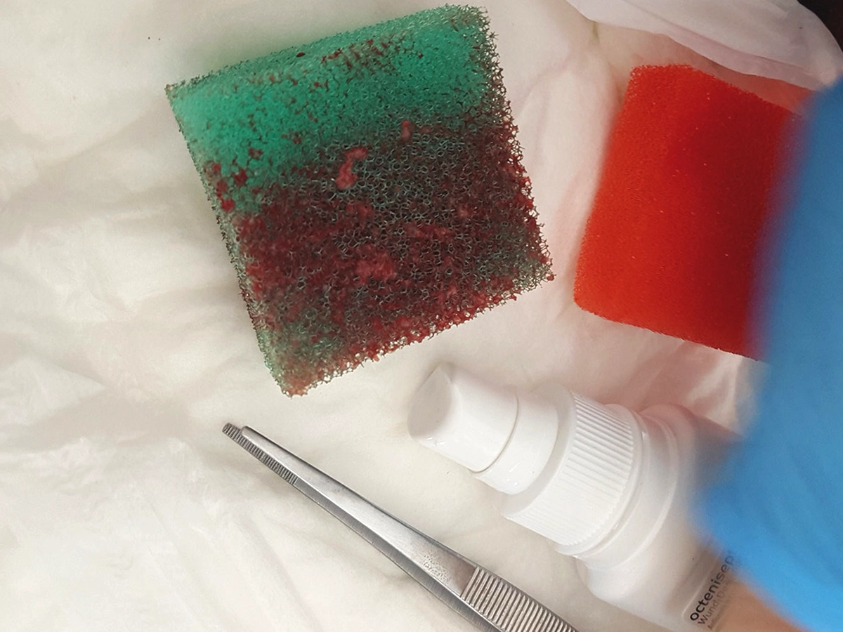
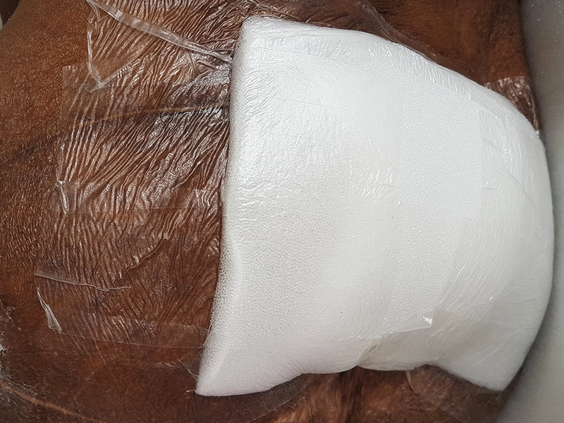
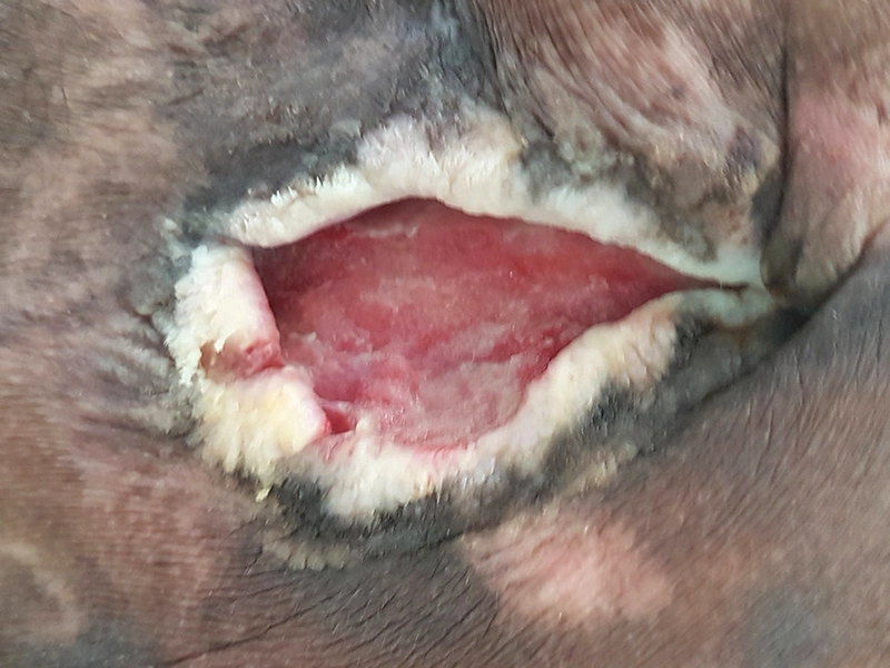
Gauze pads soaked with sterile wound irrigation solution were applied to the wound for 15 to 20 minutes. Following this, the biofilm was removed using LIGASANO® Green Scrubber wound cleansing sponges. The wound was then filled with LIGASANO® White Cavity Dressing. The primary wound dressing was LIGASANO® White Dressing, size 4 x 4 x 0.4 in (10 x 10 x 1 cm), and the secondary wound dressing was LIGASANO® Pressure Relief Foam White. The dressing was secured with Fixomull stretch and a soft fixation garment.
-
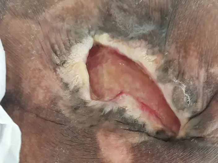
Fig. 1: April 12, 2017: wound condition before treatment with LIGASANO® White Dressing -
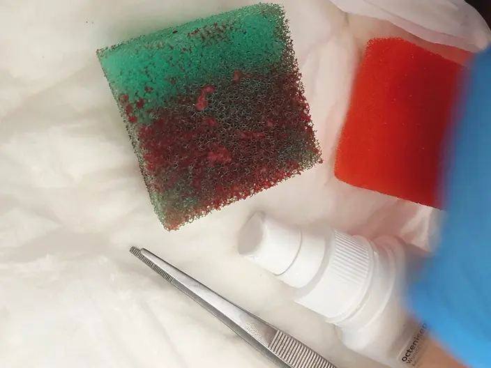
Fig. 2: April 12, 2017: mechanical debridement with LIGASANO® Green Scrubber -
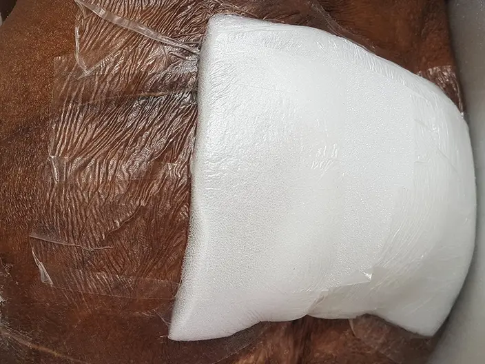
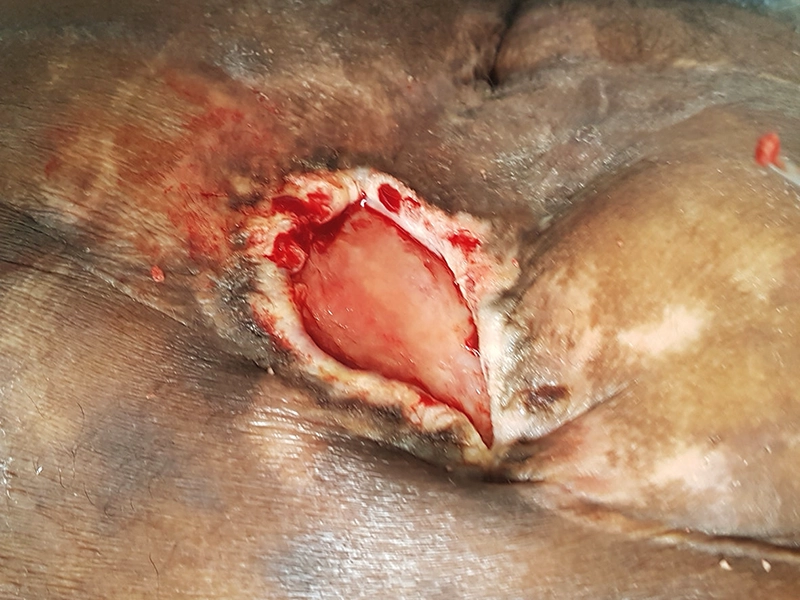
Fig. 3: April 18, 2017: the wound is covered with LIGASANO® Pressure Relief Foam for pressure reduction, and fixed with adhesive film -
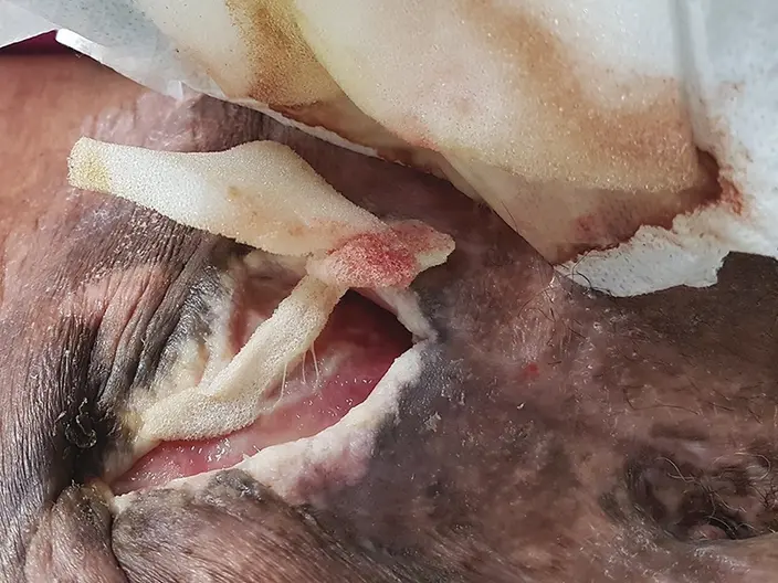
Fig. 4: April 21, 2017: the minimal undermining was packed with LIGASANO® White Dressing, and daily dressing changes were necessary -
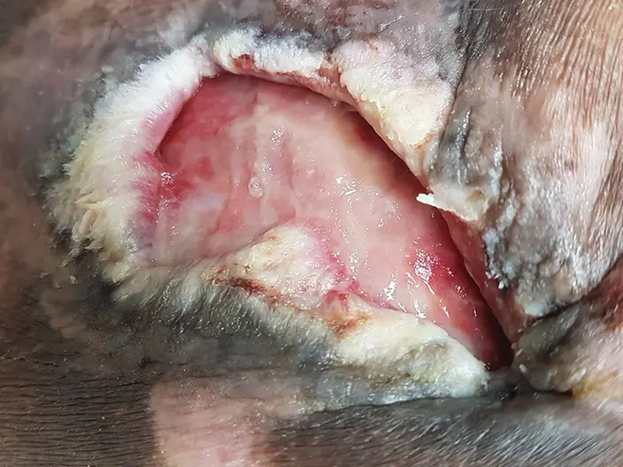
Fig. 5: April 27, 2017: the junction between the wound bed and wound edges had improved significantly, and wound depth had decreased -
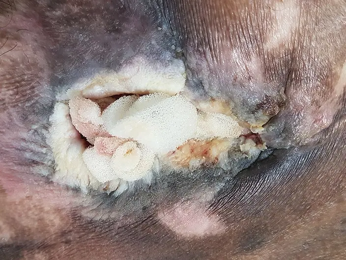
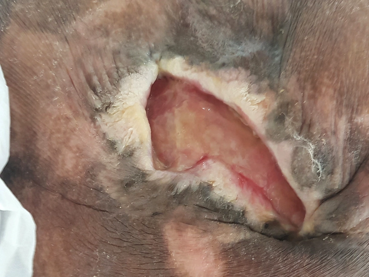
Fig. 6: May 5, 2017 the wound was completely filled with LIGASANO® White Dressing up to the edges -
-
-
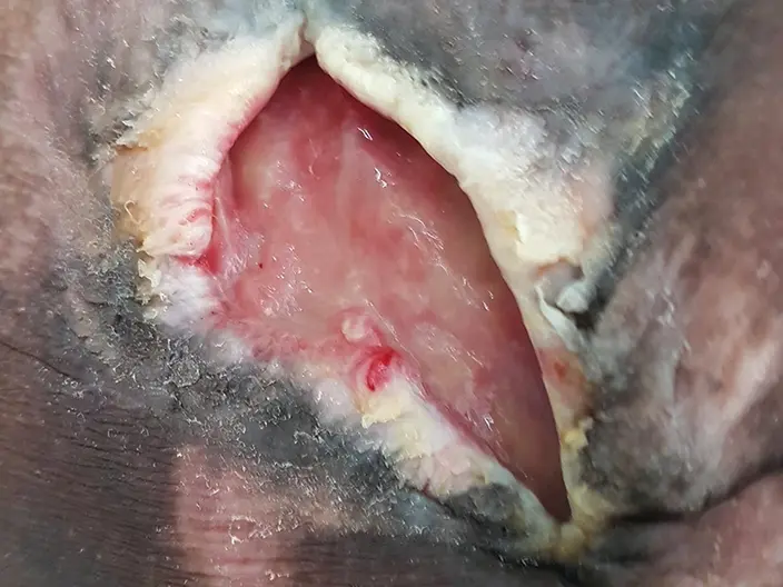
Fig. 6: May 5, 2017 the wound was completely filled with LIGASANO® White Dressing up to the edges
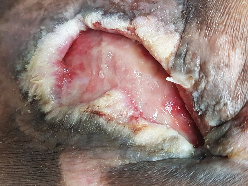
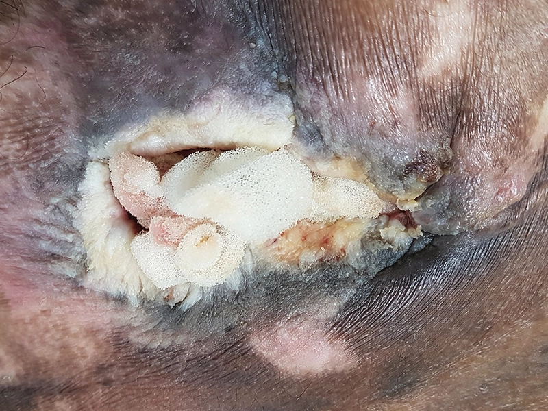
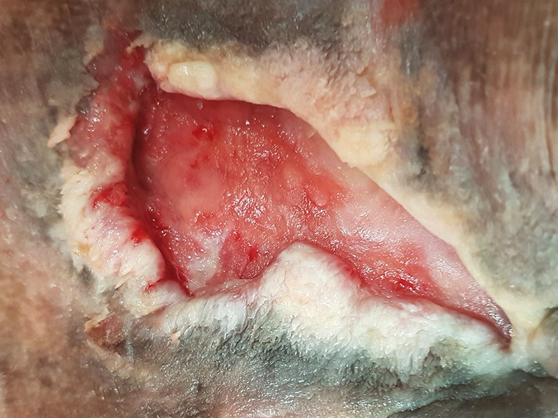
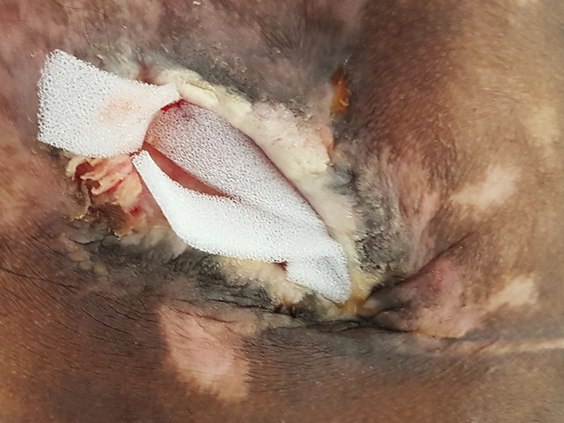
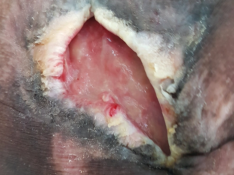
After two months of treatment with LIGASANO®, the wound has progressed very slowly toward healing, with only minor improvements observed. The patient‘s comorbidities have significantly hindered the healing process. Intensive monitoring and perseverance, as well as close collaboration with the healthcare team and the primary care physician, have been crucial to the treatment.
-
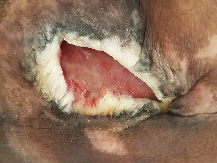
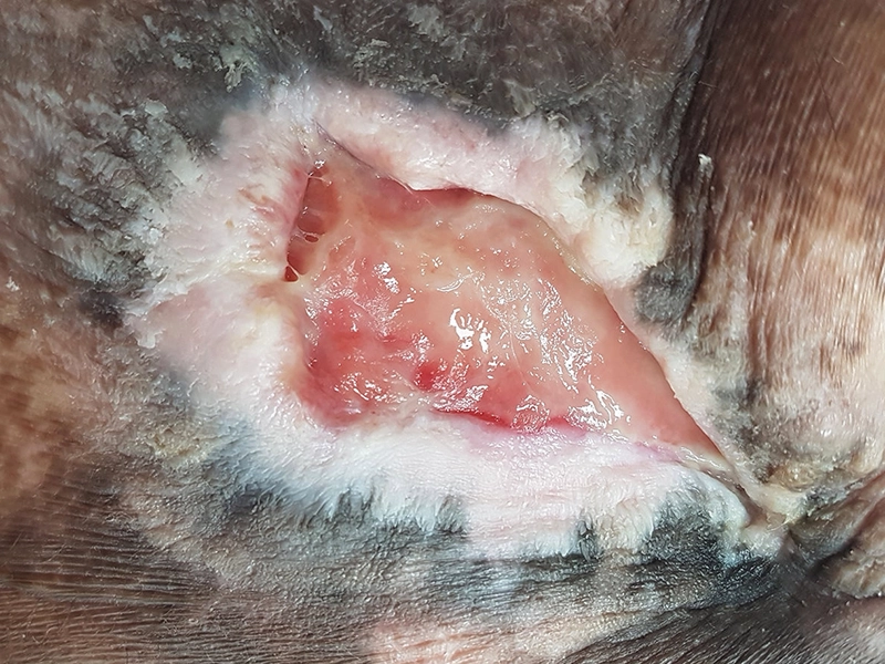
Fig. 10: on June 13, 2017 the wound edges appeared keratinized and devitalized, but they were showing increased activity in reconnecting with the wound bed. The wound itself had noticeably reduced in size and was clean. -
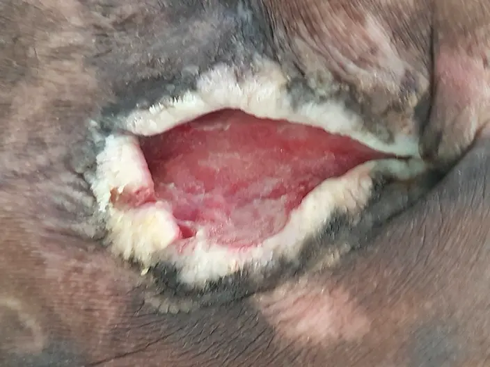
Fig. 11: on July 20, 2017, the wound continued to show improvement. However, poorly controlled diabetes and an additional wound on the trochanter were slowing down the healing process. -
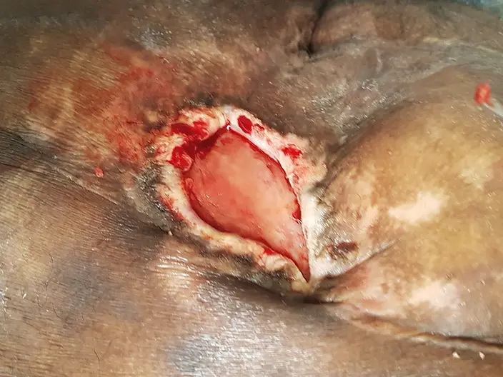
Fig. 12: on July 25, 2017, the wound condition was assessed following surgical debridement at BG Clinic Murnau in Germany -
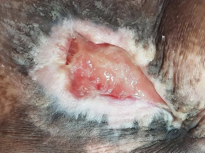
Fig. 13: September 12, 2017: despite plenty of exudate and biofilm a good junction between the wound bed and the wound edges has been developed. -
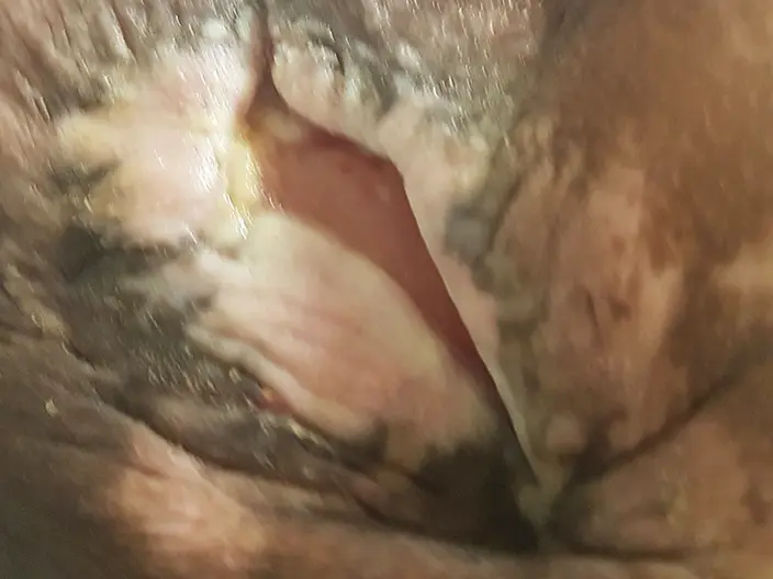
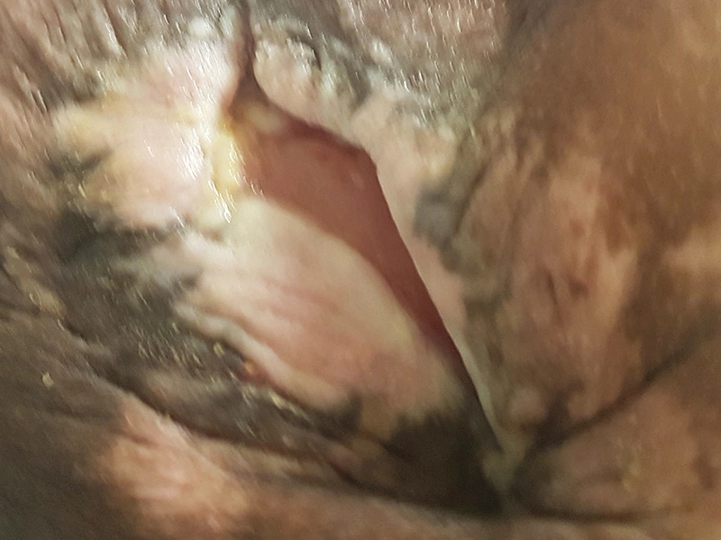
Fig. 14: on November 7, 2017, following surgery at the trochanter and with stable blood glucose levels, there was rapid progress in wound healing. The wound had reduced in size by 50%, and the wound edges were smooth, depigmented, and viable. -
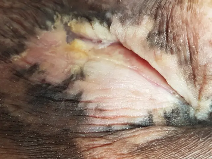
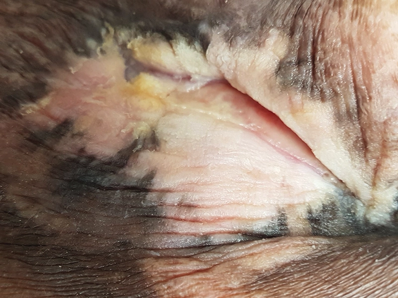
Fig. 15: on December 14, 2017, the wound was fully closed. The wound bed exhibited smooth, rosy skin formation, and the wound edges were level and well-aligned.
Conclusion:
The patient‘s overall condition was poor, with problematic kidney parameters, thyroid values, and blood glucose levels. Managing wounds in paraplegic patients presents significant challenges due to unpredictable skin temperature fluctuations, including sweating, cooling, and overheating. Complete wound closure was documented after approximately eight months.